Crystal structure of the human lamin A coil 2B dimer: implications for the head-to-tail association of nuclear lamins
Strelkov, S.V., Schumacher, J., Burkhard, P., Aebi, U., Herrmann, H.(2004) J Mol Biol 343: 1067-1080
- PubMed: 15476822
- DOI: https://doi.org/10.1016/j.jmb.2004.08.093
- Primary Citation of Related Structures:
1X8Y - PubMed Abstract:
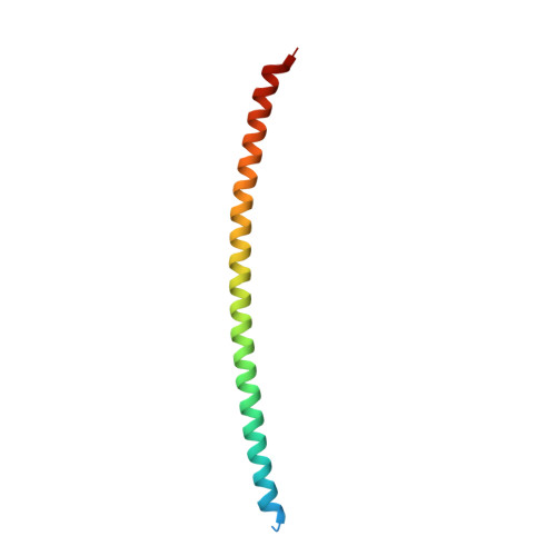
Nuclear intermediate filaments (IFs) are made from fibrous proteins termed lamins that assemble, in association with several transmembrane proteins of the inner nuclear membrane and an unknown number of chromatin proteins, into a filamentous scaffold called the nuclear lamina. In man, three types of lamins with significant sequence identity, i.e. lamin A/C, lamin B1 and B2, are expressed. The molecular characteristics of the filaments they form and the details of the assembly mechanism are still largely unknown. Here we report the crystal structure of the coiled-coil dimer from the second half of coil 2 from human lamin A at 2.2A resolution. Comparison to the recently solved structure of the homologous segment of human vimentin reveals a similar overall structure but a different distribution of charged residues and a different pattern of intra- and interhelical salt bridges. These features may explain, at least in part, the differences observed between the lamin and vimentin assembly pathways. Employing a modeled lamin A coil 1A dimer, we propose that the head-to-tail association of two lamin dimers involves strong electrostatic attractions of distinct clusters of negative charge located on the opposite ends of the rod domain with arginine clusters in the head domain and the first segment of the tail domain. Moreover, lamin A mutations, including several in coil 2B, have been associated with human laminopathies. Based on our data most of these mutations are unlikely to alter the structure of the dimer but may affect essential molecular interactions occurring in later stages of filament assembly and lamina formation.
Organizational Affiliation:
Maurice E. Müller Institute for Structural Biology, Biozentrum, University of Basel, Klingelbergstrasse 70, CH-4056 Basel, Switzerland.