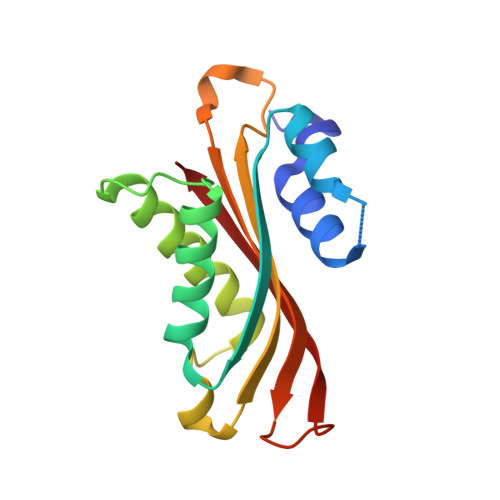
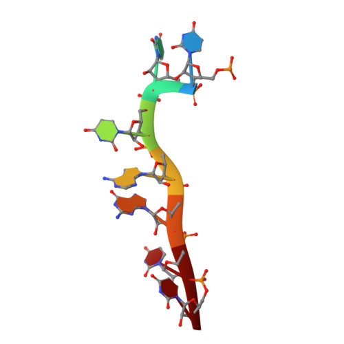
1WMQ

Structure of the HutP antitermination complex bound to a single stranded region of hut mRNA


Experimental Data Snapshot

  • Method: X-RAY DIFFRACTION
  • Resolution: 1.60 Å
  • R-Value Free: 0.249 
  • R-Value Work: 0.225 
  • R-Value Observed: 0.225 

wwPDB Validation   3D Report Full Report


Ligand Structure Quality Assessment 


This is version 1.5 of the entry. See complete history


Literature

Structural basis of HutP-mediated anti-termination and roles of the Mg2+ ion and L-histidine ligand.

Kumarevel, T.Mizuno, H.Kumar, P.K.

(2005) Nature 434: 183-191

  • DOI: https://doi.org/10.1038/nature03355
  • Primary Citation of Related Structures:  
    1WMQ, 1WPS, 1WPV

  • PubMed Abstract: 

    HutP regulates the expression of the hut structural genes of Bacillus subtilis by an anti-termination mechanism and requires two components, Mg2+ ions and L-histidine. HutP recognizes three UAG triplet units, separated by four non-conserved nucleotides on the terminator region. Here we report the 1.60-A resolution crystal structure of the quaternary complex (HutP-L-histidine-Mg2+-21-base single-stranded RNA). In the complex, the RNA adopts a novel triangular fold on the hexameric surface of HutP, without any base-pairing, and binds to the protein mostly by specific protein-base interactions. The structure explains how the HutP and RNA interactions are regulated critically by the l-histidine and Mg2+ ion through the structural rearrangement. To gain insights into these structural rearrangements, we solved two additional crystal structures (uncomplexed HutP and HutP-L-histidine-Mg2+) that revealed the intermediate structures of HutP (before forming an active structure) and the importance of the Mg2+ ion interactions in the complexes.


  • Organizational Affiliation

    Functional Nucleic Acids Group, Institute for Biological Resources and Functions, National Institute of Advanced Industrial Science and Technology (AIST), Central 6, 1-1-1 Higashi, Tsukuba, Ibaraki 305-8566, Japan.


Macromolecules

Find similar proteins by:  (by identity cutoff)  |  3D Structure
Entity ID: 2
MoleculeChains Sequence LengthOrganismDetailsImage
Hut operon positive regulatory proteinC [auth A],
D [auth B]
147Bacillus subtilisMutation(s): 1 
UniProt
Find proteins for P10943 (Bacillus subtilis (strain 168))
Explore P10943 
Go to UniProtKB:  P10943
Entity Groups  
Sequence Clusters30% Identity50% Identity70% Identity90% Identity95% Identity100% Identity
UniProt GroupP10943
Sequence Annotations
Expand
  • Reference Sequence

Find similar nucleic acids by:  Sequence   |   3D Structure  

Entity ID: 1
MoleculeChains LengthOrganismImage
5'-R(P*UP*UP*UP*AP*GP*UP*U)-3'A [auth C],
B [auth D]
7N/A
Sequence Annotations
Expand
  • Reference Sequence
Experimental Data & Validation

Experimental Data

  • Method: X-RAY DIFFRACTION
  • Resolution: 1.60 Å
  • R-Value Free: 0.249 
  • R-Value Work: 0.225 
  • R-Value Observed: 0.225 
  • Space Group: H 3
Unit Cell:
Length ( Å )Angle ( ˚ )
a = 76.21α = 90
b = 76.21β = 90
c = 133.56γ = 120
Software Package:
Software NamePurpose
CNSrefinement
HKL-2000data reduction
CCP4data scaling
MOLREPphasing
PHASERphasing

Structure Validation

View Full Validation Report


Ligand Structure Quality Assessment 


Entry History 

Deposition Data

Revision History  (Full details and data files)

  • Version 1.0: 2005-03-15
    Type: Initial release
  • Version 1.1: 2008-04-30
    Changes: Version format compliance
  • Version 1.2: 2011-07-13
    Changes: Version format compliance
  • Version 1.3: 2017-10-11
    Changes: Advisory, Refinement description
  • Version 1.4: 2021-11-10
    Changes: Advisory, Database references, Derived calculations
  • Version 1.5: 2023-10-25
    Changes: Data collection, Refinement description