1TYE

Structural basis for allostery in integrins and binding of ligand-mimetic therapeutics to the platelet receptor for fibrinogen


Experimental Data Snapshot

  • Method: X-RAY DIFFRACTION
  • Resolution: 2.90 Å
  • R-Value Free: 0.254 
  • R-Value Work: 0.219 
  • R-Value Observed: 0.268 

wwPDB Validation   3D Report Full Report


Ligand Structure Quality Assessment 


This is version 2.1 of the entry. See complete history


Literature

Structural basis for allostery in integrins and binding to fibrinogen-mimetic therapeutics

Xiao, T.Takagi, J.Coller, B.S.Wang, J.-H.Springer, T.A.

(2004) Nature 432: 59-67

  • DOI: https://doi.org/10.1038/nature02976
  • Primary Citation of Related Structures:  
    1TYE

  • PubMed Abstract: 

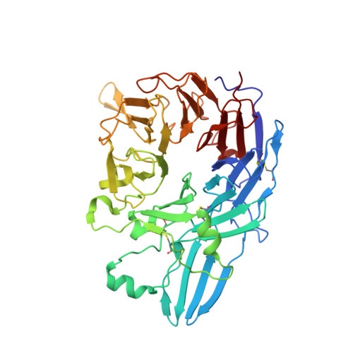
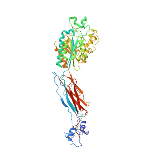
    Integrins are important adhesion receptors in all Metazoa that transmit conformational change bidirectionally across the membrane. Integrin alpha and beta subunits form a head and two long legs in the ectodomain and span the membrane. Here, we define with crystal structures the atomic basis for allosteric regulation of the conformation and affinity for ligand of the integrin ectodomain, and how fibrinogen-mimetic therapeutics bind to platelet integrin alpha(IIb)beta3. Allostery in the beta3 I domain alters three metal binding sites, associated loops and alpha1- and alpha7-helices. Piston-like displacement of the alpha7-helix causes a 62 degrees reorientation between the beta3 I and hybrid domains. Transmission through the rigidly connected plexin/semaphorin/integrin (PSI) domain in the upper beta3 leg causes a 70 A separation between the knees of the alpha and beta legs. Allostery in the head thus disrupts interaction between the legs in a previously described low-affinity bent integrin conformation, and leg extension positions the high-affinity head far above the cell surface.


  • Organizational Affiliation

    The CBR Institute for Biomedical Research and Department of Pathology, Harvard Medical School, 200 Longwood Avenue, Boston, Massachusetts 02115, USA.


Macromolecules
Find similar proteins by:  (by identity cutoff)  |  3D Structure
Entity ID: 1
MoleculeChains Sequence LengthOrganismDetailsImage
Integrin alpha-IIb
A, C, E
452Homo sapiensMutation(s): 0 
Gene Names: ITGAB
UniProt & NIH Common Fund Data Resources
Find proteins for P08514 (Homo sapiens)
Explore P08514 
Go to UniProtKB:  P08514
PHAROS:  P08514
GTEx:  ENSG00000005961 
Entity Groups  
Sequence Clusters30% Identity50% Identity70% Identity90% Identity95% Identity100% Identity
UniProt GroupP08514
Sequence Annotations
Expand
  • Reference Sequence
Find similar proteins by:  (by identity cutoff)  |  3D Structure
Entity ID: 2
MoleculeChains Sequence LengthOrganismDetailsImage
Integrin beta-3
B, D, F
440Homo sapiensMutation(s): 0 
Gene Names: ITGAB
UniProt & NIH Common Fund Data Resources
Find proteins for P05106 (Homo sapiens)
Explore P05106 
Go to UniProtKB:  P05106
PHAROS:  P05106
GTEx:  ENSG00000259207 
Entity Groups  
Sequence Clusters30% Identity50% Identity70% Identity90% Identity95% Identity100% Identity
UniProt GroupP05106
Sequence Annotations
Expand
  • Reference Sequence
Oligosaccharides

Help

Entity ID: 3
MoleculeChains Length2D Diagram Glycosylation3D Interactions
beta-D-mannopyranose-(1-3)-alpha-D-mannopyranose-(1-4)-2-acetamido-2-deoxy-beta-D-glucopyranose-(1-4)-2-acetamido-2-deoxy-beta-D-glucopyranose
G, I
4N-Glycosylation
Glycosylation Resources
GlyTouCan:  G24199JF
GlyCosmos:  G24199JF
GlyGen:  G24199JF
Entity ID: 4
MoleculeChains Length2D Diagram Glycosylation3D Interactions
beta-D-mannopyranose-(1-3)-[alpha-D-mannopyranose-(1-6)]alpha-D-mannopyranose-(1-4)-2-acetamido-2-deoxy-beta-D-glucopyranose-(1-4)-2-acetamido-2-deoxy-beta-D-glucopyranose
H
5N-Glycosylation
Glycosylation Resources
GlyTouCan:  G66753FB
GlyCosmos:  G66753FB
GlyGen:  G66753FB
Small Molecules
Ligands 4 Unique
IDChains Name / Formula / InChI Key2D Diagram3D Interactions
NAG
Query on NAG

Download Ideal Coordinates CCD File 
DA [auth E]
IA [auth F]
JA [auth F]
N [auth B]
O [auth B]
DA [auth E],
IA [auth F],
JA [auth F],
N [auth B],
O [auth B],
X [auth D],
Y [auth D]
2-acetamido-2-deoxy-beta-D-glucopyranose
C8 H15 N O6
OVRNDRQMDRJTHS-FMDGEEDCSA-N
CAC
Query on CAC

Download Ideal Coordinates CCD File 
KA [auth F],
P [auth B],
Z [auth D]
CACODYLATE ION
C2 H6 As O2
OGGXGZAMXPVRFZ-UHFFFAOYSA-M
CA
Query on CA

Download Ideal Coordinates CCD File 
BA [auth D]
CA [auth D]
EA [auth E]
FA [auth E]
GA [auth E]
BA [auth D],
CA [auth D],
EA [auth E],
FA [auth E],
GA [auth E],
HA [auth E],
J [auth A],
K [auth A],
L [auth A],
M [auth A],
MA [auth F],
NA [auth F],
R [auth B],
S [auth B],
T [auth C],
U [auth C],
V [auth C],
W [auth C]
CALCIUM ION
Ca
BHPQYMZQTOCNFJ-UHFFFAOYSA-N
MG
Query on MG

Download Ideal Coordinates CCD File 
AA [auth D],
LA [auth F],
Q [auth B]
MAGNESIUM ION
Mg
JLVVSXFLKOJNIY-UHFFFAOYSA-N
Experimental Data & Validation

Experimental Data

  • Method: X-RAY DIFFRACTION
  • Resolution: 2.90 Å
  • R-Value Free: 0.254 
  • R-Value Work: 0.219 
  • R-Value Observed: 0.268 
  • Space Group: P 62
Unit Cell:
Length ( Å )Angle ( ˚ )
a = 332.093α = 90
b = 332.093β = 90
c = 88.288γ = 120
Software Package:
Software NamePurpose
HKL-2000data collection
SCALEPACKdata scaling
AMoREphasing
CNSrefinement
HKL-2000data reduction

Structure Validation

View Full Validation Report


Ligand Structure Quality Assessment 


Entry History 

Deposition Data

Revision History  (Full details and data files)

  • Version 1.0: 2004-10-12
    Type: Initial release
  • Version 1.1: 2008-04-30
    Changes: Version format compliance
  • Version 1.2: 2011-07-13
    Changes: Non-polymer description, Version format compliance
  • Version 1.3: 2014-09-10
    Changes: Database references
  • Version 2.0: 2020-07-29
    Type: Remediation
    Reason: Carbohydrate remediation
    Changes: Atomic model, Data collection, Derived calculations, Structure summary
  • Version 2.1: 2023-08-23
    Changes: Data collection, Database references, Refinement description, Structure summary