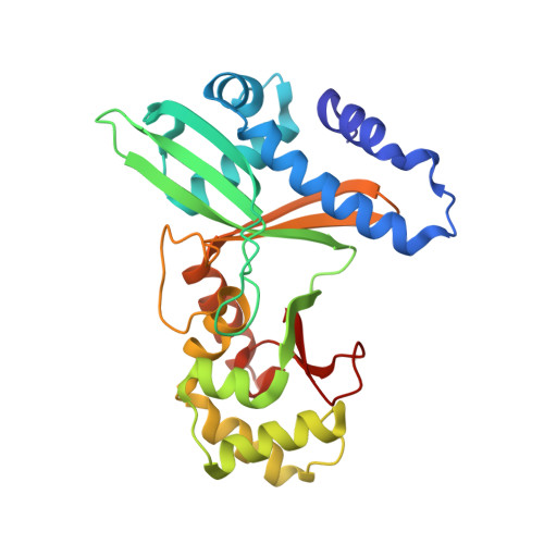
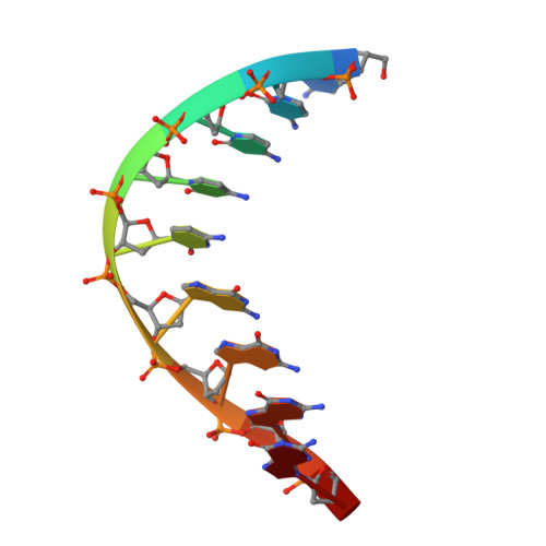
1SA3

An asymmetric complex of restriction endonuclease MspI on its palindromic DNA recognition site


Experimental Data Snapshot

  • Method: X-RAY DIFFRACTION
  • Resolution: 1.95 Å
  • R-Value Free: 0.252 
  • R-Value Work: 0.223 
  • R-Value Observed: 0.225 

wwPDB Validation   3D Report Full Report


This is version 1.4 of the entry. See complete history


Literature

An Asymmetric Complex of Restriction Endonuclease MspI on Its Palindromic DNA Recognition Site.

Xu, Q.S.Kucera, R.B.Roberts, R.J.Guo, H.C.

(2004) Structure 12: 1741-1747

  • DOI: https://doi.org/10.1016/j.str.2004.07.014
  • Primary Citation of Related Structures:  
    1SA3

  • PubMed Abstract: 

    Most well-known restriction endonucleases recognize palindromic DNA sequences and are classified as Type IIP. Due to the recognition and cleavage symmetry, Type IIP enzymes are usually found to act as homodimers in forming 2-fold symmetric enzyme-DNA complexes. Here we report an asymmetric complex of the Type IIP restriction enzyme MspI in complex with its cognate recognition sequence. Unlike any other Type IIP enzyme reported to date, an MspI monomer and not a dimer binds to a palindromic DNA sequence. The enzyme makes specific contacts with all 4 base pairs in the recognition sequence, by six direct and five water-mediated hydrogen bonds and numerous van der Waal contacts. This MspI-DNA structure represents the first example of asymmetric recognition of a palindromic DNA sequence by two different structural motifs in one polypeptide. A few possible pathways are discussed for MspI to cut both strands of DNA, either as a monomer or dimer.


  • Organizational Affiliation

    Department of Physiology and Biophysics, Boston University School of Medicine, 715 Albany Street, MA 02118, USA.


Macromolecules

Find similar proteins by:  (by identity cutoff)  |  3D Structure
Entity ID: 2
MoleculeChains Sequence LengthOrganismDetailsImage
Type II restriction enzyme MspIE [auth A],
F [auth B]
262Moraxella sp.Mutation(s): 0 
Gene Names: MSPIR
EC: 3.1.21.4
UniProt
Find proteins for P11405 (Moraxella sp)
Explore P11405 
Go to UniProtKB:  P11405
Entity Groups  
Sequence Clusters30% Identity50% Identity70% Identity90% Identity95% Identity100% Identity
UniProt GroupP11405
Sequence Annotations
Expand
  • Reference Sequence

Find similar nucleic acids by:  Sequence   |   3D Structure  

Entity ID: 1
MoleculeChains LengthOrganismImage
5'-D(*CP*CP*CP*CP*CP*GP*GP*GP*GP*G)-3'A [auth C],
B [auth D],
C [auth E],
D [auth F]
10N/A
Sequence Annotations
Expand
  • Reference Sequence
Experimental Data & Validation

Experimental Data

  • Method: X-RAY DIFFRACTION
  • Resolution: 1.95 Å
  • R-Value Free: 0.252 
  • R-Value Work: 0.223 
  • R-Value Observed: 0.225 
  • Space Group: P 1 21 1
Unit Cell:
Length ( Å )Angle ( ˚ )
a = 50.193α = 90
b = 131.601β = 109.69
c = 59.318γ = 90
Software Package:
Software NamePurpose
CNSrefinement
DENZOdata reduction
SCALEPACKdata scaling
DMphasing

Structure Validation

View Full Validation Report


Entry History 

Deposition Data

Revision History  (Full details and data files)

  • Version 1.0: 2004-09-07
    Type: Initial release
  • Version 1.1: 2008-04-29
    Changes: Version format compliance
  • Version 1.2: 2011-07-13
    Changes: Version format compliance
  • Version 1.3: 2018-01-31
    Changes: Advisory, Experimental preparation
  • Version 1.4: 2024-02-14
    Changes: Advisory, Data collection, Database references, Derived calculations