1S78

Insights into ErbB signaling from the structure of the ErbB2-pertuzumab complex


Experimental Data Snapshot

  • Method: X-RAY DIFFRACTION
  • Resolution: 3.25 Å
  • R-Value Free: 0.268 
  • R-Value Work: 0.224 
  • R-Value Observed: 0.227 

wwPDB Validation   3D Report Full Report


Ligand Structure Quality Assessment 


This is version 2.1 of the entry. See complete history


Literature

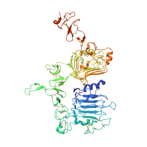
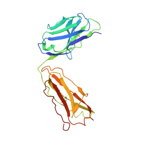
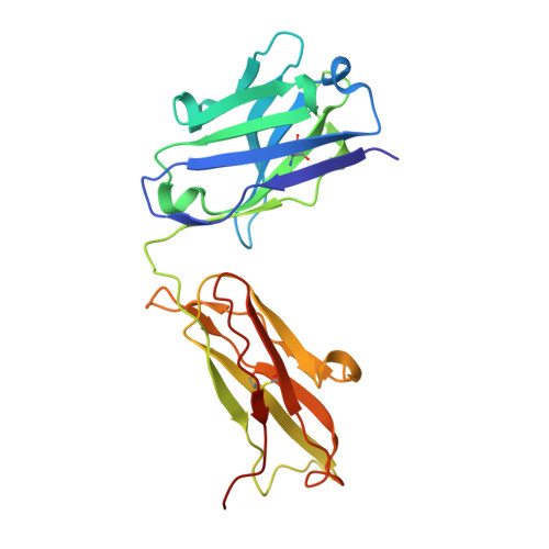
Insights into ErbB signaling from the structure of the ErbB2-pertuzumab complex.

Franklin, M.C.Carey, K.D.Vajdos, F.F.Leahy, D.J.De Vos, A.M.Sliwkowski, M.X.

(2004) Cancer Cell 5: 317-328

  • DOI: https://doi.org/10.1016/s1535-6108(04)00083-2
  • Primary Citation of Related Structures:  
    1S78

  • PubMed Abstract: 

    We have determined the 3.2 A X-ray crystal structure of the extracellular domain of the human epidermal growth factor receptor 2 (ErbB2 or HER2) in a complex with the antigen binding fragment of pertuzumab, an anti-ErbB2 monoclonal antibody also known as 2C4 or Omnitarg. Pertuzumab binds to ErbB2 near the center of domain II, sterically blocking a binding pocket necessary for receptor dimerization and signaling. The ErbB2-pertuzumab structure, combined with earlier mutagenesis data, defines the pertuzumab residues essential for ErbB2 interaction. To analyze the ErbB2 side of the interface, we have mutated a number of residues contacting pertuzumab and examined the effects of these mutations on pertuzumab binding and ErbB2-ErbB3 heterodimerization. We have also shown that conserved residues previously shown to be necessary for EGF receptor homodimerization may be dispensible for ErbB2-ErbB3 heterodimerization.


  • Organizational Affiliation

    Department of Protein Engineering, Genentech, Inc., 1 DNA Way, South San Francisco, CA 94114 USA.


Macromolecules
Find similar proteins by:  (by identity cutoff)  |  3D Structure
Entity ID: 1
MoleculeChains Sequence LengthOrganismDetailsImage
Receptor protein-tyrosine kinase erbB-2
A, B
624Homo sapiensMutation(s): 0 
Gene Names: ERBB2HER2NGLNEU
EC: 2.7.1.112
UniProt & NIH Common Fund Data Resources
Find proteins for P04626 (Homo sapiens)
Explore P04626 
Go to UniProtKB:  P04626
PHAROS:  P04626
GTEx:  ENSG00000141736 
Entity Groups  
Sequence Clusters30% Identity50% Identity70% Identity90% Identity95% Identity100% Identity
UniProt GroupP04626
Sequence Annotations
Expand
  • Reference Sequence
Find similar proteins by:  (by identity cutoff)  |  3D Structure
Entity ID: 2
MoleculeChains Sequence LengthOrganismDetailsImage
Pertuzumab Fab light chain
C, E
214Mus musculusMutation(s): 0 
UniProt
Find proteins for Q7Z3Y4 (Homo sapiens)
Explore Q7Z3Y4 
Go to UniProtKB:  Q7Z3Y4
Entity Groups  
Sequence Clusters30% Identity50% Identity70% Identity90% Identity95% Identity100% Identity
UniProt GroupQ7Z3Y4
Sequence Annotations
Expand
  • Reference Sequence
Find similar proteins by:  (by identity cutoff)  |  3D Structure
Entity ID: 3
MoleculeChains Sequence LengthOrganismDetailsImage
Pertuzumab Fab heavy chain
D, F
226Mus musculusMutation(s): 0 
Entity Groups  
Sequence Clusters30% Identity50% Identity70% Identity90% Identity95% Identity100% Identity
Sequence Annotations
Expand
  • Reference Sequence
Oligosaccharides

Help

Entity ID: 4
MoleculeChains Length2D Diagram Glycosylation3D Interactions
2-acetamido-2-deoxy-beta-D-glucopyranose-(1-4)-2-acetamido-2-deoxy-beta-D-glucopyranose
G, H, I, K
2N-Glycosylation
Glycosylation Resources
GlyTouCan:  G42666HT
GlyCosmos:  G42666HT
GlyGen:  G42666HT
Entity ID: 5
MoleculeChains Length2D Diagram Glycosylation3D Interactions
beta-D-mannopyranose-(1-4)-2-acetamido-2-deoxy-beta-D-glucopyranose-(1-4)-2-acetamido-2-deoxy-beta-D-glucopyranose
J
3N-Glycosylation
Glycosylation Resources
GlyTouCan:  G15407YE
GlyCosmos:  G15407YE
GlyGen:  G15407YE
Small Molecules
Ligands 1 Unique
IDChains Name / Formula / InChI Key2D Diagram3D Interactions
NAG
Query on NAG

Download Ideal Coordinates CCD File 
L [auth A],
M [auth B]
2-acetamido-2-deoxy-beta-D-glucopyranose
C8 H15 N O6
OVRNDRQMDRJTHS-FMDGEEDCSA-N
Experimental Data & Validation

Experimental Data

  • Method: X-RAY DIFFRACTION
  • Resolution: 3.25 Å
  • R-Value Free: 0.268 
  • R-Value Work: 0.224 
  • R-Value Observed: 0.227 
  • Space Group: P 43 21 2
Unit Cell:
Length ( Å )Angle ( ˚ )
a = 139.412α = 90
b = 139.412β = 90
c = 356.873γ = 90
Software Package:
Software NamePurpose
REFMACrefinement
DENZOdata reduction
SCALEPACKdata scaling
AMoREphasing

Structure Validation

View Full Validation Report


Ligand Structure Quality Assessment 


Entry History 

Deposition Data

Revision History  (Full details and data files)

  • Version 1.0: 2004-04-27
    Type: Initial release
  • Version 1.1: 2008-04-29
    Changes: Version format compliance
  • Version 1.2: 2011-07-13
    Changes: Advisory, Version format compliance
  • Version 2.0: 2020-07-29
    Type: Remediation
    Reason: Carbohydrate remediation
    Changes: Advisory, Atomic model, Data collection, Derived calculations, Structure summary
  • Version 2.1: 2023-08-23
    Changes: Data collection, Database references, Refinement description, Structure summary