1S0Z

Crystal structure of the VDR LBD complexed to seocalcitol.


Experimental Data Snapshot

  • Method: X-RAY DIFFRACTION
  • Resolution: 2.50 Å
  • R-Value Free: 0.204 
  • R-Value Work: 0.170 
  • R-Value Observed: 0.204 

wwPDB Validation   3D Report Full Report


Ligand Structure Quality Assessment 


This is version 1.4 of the entry. See complete history


Literature

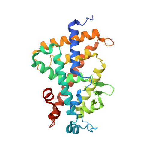
Crystal structures of the vitamin D nuclear receptor liganded with the vitamin D side chain analogues calcipotriol and seocalcitol, receptor agonists of clinical importance. Insights into a structural basis for the switching of calcipotriol to a receptor antagonist by further side chain modification.

Tocchini-Valentini, G.Rochel, N.Wurtz, J.M.Moras, D.

(2004) J Med Chem 47: 1956-1961

  • DOI: https://doi.org/10.1021/jm0310582
  • Primary Citation of Related Structures:  
    1S0Z, 1S19

  • PubMed Abstract: 

    The plethora of actions of 1alpha,25(OH)(2)D(3) in various systems suggested wide clinical applications of vitamin D nuclear receptor (VDR) ligands in treatments of inflammation, dermatological indication, osteoporosis, cancers, and autoimmune diseases. More than 3000 vitamin D analogues have been synthesized in order to reduce the calcemic side effects while maintaining the transactivation potency of these ligands. Here, we report the crystal structures of VDR ligand binding domain bound to two vitamin D agonists of therapeutical interest, calcipotriol and seocalcitol, which are characterized by their side chain modifications. These structures show the conservation of the VDR structure and the adaptation of the side chain anchored by hydroxyl moieties. The structure of VDR-calcipotriol helps us to understand the structural basis for for the switching of calcipotriol to a receptor antagonist by further side chain modification. The VDR-seocalcitol structure, in comparison with the structure of VDR-KH1060, a superagonist ligand closely related to seocalcitol, shows adaptation of the D ring and position of C-21 in order to adapt its more rigid side chain.


  • Organizational Affiliation

    Département de Biologie et de Génomique Structurales, IGBMC, CNRS/INSERM/Université Louis Pasteur, Parc d'Innovation BP10142, 67404 Illkirch Cedex, France.


Macromolecules
Find similar proteins by:  (by identity cutoff)  |  3D Structure
Entity ID: 1
MoleculeChains Sequence LengthOrganismDetailsImage
Vitamin D3 receptor263Homo sapiensMutation(s): 0 
Gene Names: VDRNR1I1
UniProt & NIH Common Fund Data Resources
Find proteins for P11473 (Homo sapiens)
Explore P11473 
Go to UniProtKB:  P11473
PHAROS:  P11473
GTEx:  ENSG00000111424 
Entity Groups  
Sequence Clusters30% Identity50% Identity70% Identity90% Identity95% Identity100% Identity
UniProt GroupP11473
Sequence Annotations
Expand
  • Reference Sequence
Small Molecules
Ligands 1 Unique
IDChains Name / Formula / InChI Key2D Diagram3D Interactions
EB1
Query on EB1

Download Ideal Coordinates CCD File 
B [auth A]SEOCALCITOL
C30 H46 O3
LVLLALCJVJNGQQ-SEODYNFXSA-N
Experimental Data & Validation

Experimental Data

  • Method: X-RAY DIFFRACTION
  • Resolution: 2.50 Å
  • R-Value Free: 0.204 
  • R-Value Work: 0.170 
  • R-Value Observed: 0.204 
  • Space Group: P 21 21 21
Unit Cell:
Length ( Å )Angle ( ˚ )
a = 45.134α = 90
b = 52.528β = 90
c = 132.648γ = 90
Software Package:
Software NamePurpose
HKL-2000data collection
SCALEPACKdata scaling
CNSrefinement
HKL-2000data reduction
CNSphasing

Structure Validation

View Full Validation Report


Ligand Structure Quality Assessment 


Entry History 

Deposition Data

Revision History  (Full details and data files)

  • Version 1.0: 2004-04-13
    Type: Initial release
  • Version 1.1: 2008-04-29
    Changes: Version format compliance
  • Version 1.2: 2011-07-13
    Changes: Version format compliance
  • Version 1.3: 2017-08-09
    Changes: Advisory, Refinement description, Source and taxonomy
  • Version 1.4: 2024-02-14
    Changes: Advisory, Data collection, Database references, Derived calculations, Structure summary