1QF8

TRUNCATED FORM OF CASEIN KINASE II BETA SUBUNIT (2-182) FROM HOMO SAPIENS


Experimental Data Snapshot

  • Method: X-RAY DIFFRACTION
  • Resolution: 1.74 Å
  • R-Value Free: 0.219 
  • R-Value Work: 0.194 

wwPDB Validation   3D Report Full Report


This is version 1.4 of the entry. See complete history


Literature

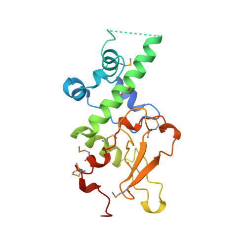
Crystal structure of the human protein kinase CK2 regulatory subunit reveals its zinc finger-mediated dimerization.

Chantalat, L.Leroy, D.Filhol, O.Nueda, A.Benitez, M.J.Chambaz, E.M.Cochet, C.Dideberg, O.

(1999) EMBO J 18: 2930-2940

  • DOI: https://doi.org/10.1093/emboj/18.11.2930
  • Primary Citation of Related Structures:  
    1QF8

  • PubMed Abstract: 

    Protein kinase CK2 is a tetramer composed of two alpha catalytic subunits and two beta regulatory subunits. The structure of a C-terminal truncated form of the human beta subunit has been determined by X-ray crystallography to 1.7 A resolution. One dimer is observed in the asymmetric unit of the crystal. The most striking feature of the structure is the presence of a zinc finger mediating the dimerization. The monomer structure consists of two domains, one entirely alpha-helical and one including the zinc finger. The dimer has a crescent shape holding a highly acidic region at both ends. We propose that this acidic region is involved in the interactions with the polyamines and/or catalytic subunits. Interestingly, conserved amino acid residues among beta subunit sequences are clustered along one linear ridge that wraps around the entire dimer. This feature suggests that protein partners may interact with the dimer through a stretch of residues in an extended conformation.


  • Organizational Affiliation

    Laboratoire de Cristallographie Macromoléculaire, Institut de Biologie Structurale Jean-Pierre Ebel, CNRS/CEA, 41, rue Jules Horowitz, 38027 Grenoble Cedex 1, France.


Macromolecules
Find similar proteins by:  (by identity cutoff)  |  3D Structure
Entity ID: 1
MoleculeChains Sequence LengthOrganismDetailsImage
CASEIN KINASE II
A, B
182Homo sapiensMutation(s): 8 
EC: 2.7.1.37
UniProt & NIH Common Fund Data Resources
Find proteins for P67870 (Homo sapiens)
Explore P67870 
Go to UniProtKB:  P67870
PHAROS:  P67870
GTEx:  ENSG00000204435 
Entity Groups  
Sequence Clusters30% Identity50% Identity70% Identity90% Identity95% Identity100% Identity
UniProt GroupP67870
Sequence Annotations
Expand
  • Reference Sequence
Small Molecules
Ligands 2 Unique
IDChains Name / Formula / InChI Key2D Diagram3D Interactions
ZN
Query on ZN

Download Ideal Coordinates CCD File 
C [auth A],
E [auth B]
ZINC ION
Zn
PTFCDOFLOPIGGS-UHFFFAOYSA-N
MG
Query on MG

Download Ideal Coordinates CCD File 
D [auth A]MAGNESIUM ION
Mg
JLVVSXFLKOJNIY-UHFFFAOYSA-N
Modified Residues  1 Unique
IDChains TypeFormula2D DiagramParent
MSE
Query on MSE
A, B
L-PEPTIDE LINKINGC5 H11 N O2 SeMET
Experimental Data & Validation

Experimental Data

  • Method: X-RAY DIFFRACTION
  • Resolution: 1.74 Å
  • R-Value Free: 0.219 
  • R-Value Work: 0.194 
  • Space Group: P 42 21 2
Unit Cell:
Length ( Å )Angle ( ˚ )
a = 132.23α = 90
b = 132.23β = 90
c = 63.78γ = 90
Software Package:
Software NamePurpose
MLPHAREphasing
SOLOMONphasing
CNSrefinement
DENZOdata reduction
CCP4data scaling
SCALAdata scaling
TRUNCATEdata scaling

Structure Validation

View Full Validation Report


Entry History 

Deposition Data

Revision History  (Full details and data files)

  • Version 1.0: 2000-04-19
    Type: Initial release
  • Version 1.1: 2007-10-16
    Changes: Version format compliance
  • Version 1.2: 2011-07-13
    Changes: Version format compliance
  • Version 1.3: 2017-10-04
    Changes: Advisory, Refinement description
  • Version 1.4: 2023-12-27
    Changes: Advisory, Data collection, Database references, Derived calculations