1PZU

An asymmetric NFAT1-RHR homodimer on a pseudo-palindromic, Kappa-B site


Experimental Data Snapshot

  • Method: X-RAY DIFFRACTION
  • Resolution: 3.10 Å
  • R-Value Free: 0.319 
  • R-Value Work: 0.296 
  • R-Value Observed: 0.303 

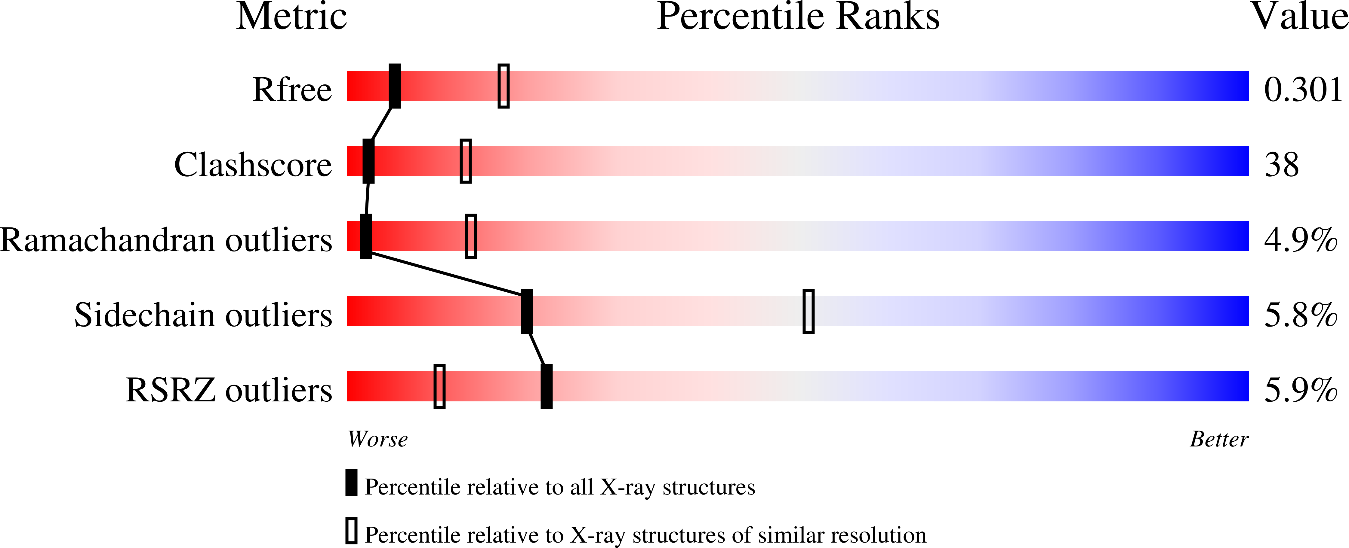
wwPDB Validation   3D Report Full Report


This is version 1.3 of the entry. See complete history


Literature

An asymmetric NFAT1 dimer on a pseudo-palindromic KB-like DNA site

Jin, L.Sliz, P.Chen, L.Macian, F.Rao, A.Hogan, P.G.Harrison, S.C.

(2003) Nat Struct Biol 10: 807-811

  • DOI: https://doi.org/10.1038/nsb975
  • Primary Citation of Related Structures:  
    1PZU

  • PubMed Abstract: 

    The crystal structure of the NFAT1 Rel homology region (RHR) bound to a pseudo-palindromic DNA site reveals an asymmetric dimer interaction between the RHR-C domains, unrelated to the contact seen in Rel dimers such as NF kappa B. Binding studies with a form of the NFAT1 RHR defective in the dimer contact show loss of cooperativity and demonstrate that the same interaction is present in solution. The structure we have determined may correspond to a functional NFAT binding mode at palindromic sites of genes induced during the anergic response to weak TCR signaling.


  • Organizational Affiliation

    Department of Biological Chemistry and Molecular Pharmacology and Howard Hughes Medical Institute, Harvard Medical School, Boston, Massachusetts 02115, USA.


Macromolecules

Find similar proteins by:  (by identity cutoff)  |  3D Structure
Entity ID: 3
MoleculeChains Sequence LengthOrganismDetailsImage
Nuclear factor of activated T-cells, cytoplasmic 2301Homo sapiensMutation(s): 0 
Gene Names: NFAT1
UniProt & NIH Common Fund Data Resources
Find proteins for Q13469 (Homo sapiens)
Explore Q13469 
Go to UniProtKB:  Q13469
PHAROS:  Q13469
GTEx:  ENSG00000101096 
Entity Groups  
Sequence Clusters30% Identity50% Identity70% Identity90% Identity95% Identity100% Identity
UniProt GroupQ13469
Sequence Annotations
Expand
  • Reference Sequence

Find similar nucleic acids by:  Sequence   |   3D Structure  

Entity ID: 1
MoleculeChains LengthOrganismImage
5'-D(*TP*TP*GP*AP*GP*GP*AP*AP*TP*TP*TP*CP*CP*A)-3'A [auth X],
C [auth V],
E [auth T]
14N/A
Sequence Annotations
Expand
  • Reference Sequence

Find similar nucleic acids by:  Sequence   |   3D Structure  

Entity ID: 2
MoleculeChains LengthOrganismImage
5'-D(*AP*AP*TP*GP*GP*AP*AP*AP*TP*TP*CP*CP*TP*C)-3'B [auth Y],
D [auth Z],
F [auth W]
14N/A
Sequence Annotations
Expand
  • Reference Sequence
Experimental Data & Validation

Experimental Data

  • Method: X-RAY DIFFRACTION
  • Resolution: 3.10 Å
  • R-Value Free: 0.319 
  • R-Value Work: 0.296 
  • R-Value Observed: 0.303 
  • Space Group: P 21 21 21
Unit Cell:
Length ( Å )Angle ( ˚ )
a = 81.7α = 90
b = 122.9β = 90
c = 241.6γ = 90
Software Package:
Software NamePurpose
CNSrefinement
DENZOdata reduction
SCALEPACKdata scaling
CNSphasing

Structure Validation

View Full Validation Report


Entry History 

Deposition Data

Revision History  (Full details and data files)

  • Version 1.0: 2003-09-09
    Type: Initial release
  • Version 1.1: 2008-04-29
    Changes: Version format compliance
  • Version 1.2: 2011-07-13
    Changes: Version format compliance
  • Version 1.3: 2024-02-21
    Changes: Data collection, Database references