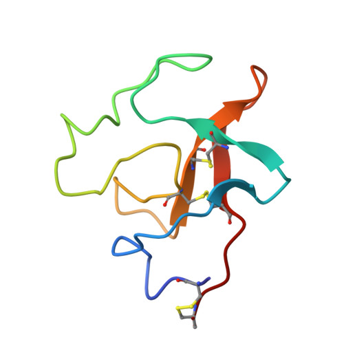
Crystal and molecular structure of human plasminogen kringle 4 refined at 1.9-A resolution.
Mulichak, A.M., Tulinsky, A., Ravichandran, K.G.(1991) Biochemistry 30: 10576-10588
- PubMed: 1657148
- DOI: https://doi.org/10.1021/bi00107a029
- Primary Citation of Related Structures:
1PK4 - PubMed Abstract:
The crystal structure of human plasminogen kringle 4 (PGK4) has been solved by molecular replacement using the bovine prothrombin kringle 1 (PTK1) structure as a model and refined by restrained least-squares methods to an R factor of 14.2% at 1.9-A resolution. The K4 structure is similar to that of PTK1, and an insertion of one residue at position 59 of the latter has minimal effect on the protein folding. The PGK4 structure is highly stabilized by an internal hydrophobic core and an extensive hydrogen-bonding network. Features new to this kringle include a cis peptide bond at Pro30 and the presence of two alternate, perpendicular, and equally occupied orientations for the Cys75 side chain. The K4 lysine-binding site consists of a hydrophobic trough formed by the Trp62 and Trp72 indole rings, with anionic (Asp55/Asp57) and cationic (Lys35/Arg71) charge pairs at either end. With the adjacent Asp5 and Arg32 residues, these result in triply charged anionic and cationic clusters (pH of crystals at 6.0), which, in addition to the unusually high accessibility of the Trp72 side chain, serve as an obvious marker of the binding site on the K4 surface. A complex intermolecular interaction occurs between the binding sites of symmetry-related molecules involving a highly ordered sulfate anion of solvation in which the Arg32 side chain of a neighboring kringle occupies the binding site.
Organizational Affiliation:
Department of Chemistry, Michigan State University, East Lansing 48824.