1OUK

The structure of p38 alpha in complex with a pyridinylimidazole inhibitor


Experimental Data Snapshot

  • Method: X-RAY DIFFRACTION
  • Resolution: 2.50 Å
  • R-Value Free: 0.264 
  • R-Value Work: 0.213 
  • R-Value Observed: 0.217 

wwPDB Validation   3D Report Full Report


Ligand Structure Quality Assessment 


This is version 1.4 of the entry. See complete history


Literature

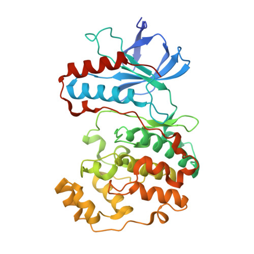
Structural basis for p38alpha MAP kinase quinazolinone and pyridol-pyrimidine inhibitor specificity

Fitzgerald, C.E.Patel, S.B.Becker, J.W.Cameron, P.M.Zaller, D.Pikounis, V.B.O'Keefe, S.J.Scapin, G.

(2003) Nat Struct Biol 10: 764-769

  • DOI: https://doi.org/10.1038/nsb949
  • Primary Citation of Related Structures:  
    1OUK, 1OUY, 1OVE

  • PubMed Abstract: 

    The quinazolinone and pyridol-pyrimidine classes of p38 MAP kinase inhibitors have a previously unseen degree of specificity for p38 over other MAP kinases. Comparison of the crystal structures of p38 bound to four different compounds shows that binding of the more specific molecules is characterized by a peptide flip between Met109 and Gly110. Gly110 is a residue specific to the alpha, beta and gamma isoforms of p38. The delta isoform and the other MAP kinases have bulkier residues in this position. These residues would likely make the peptide flip energetically unfavorable, thus explaining the selectivity of binding. To test this hypothesis, we constructed G110A and G110D mutants of p38 and measured the potency of several compounds against them. The results confirm that the selectivity of quinazolinones and pyridol-pyrimidines results from the presence of a glycine in position 110. This unique mode of binding may be exploited in the design of new p38 inhibitors.


  • Organizational Affiliation

    Department of Immunology, Merck Research Laboratories, PO Box 2000, Rahway, New Jersey 07065, USA.


Macromolecules
Find similar proteins by:  (by identity cutoff)  |  3D Structure
Entity ID: 1
MoleculeChains Sequence LengthOrganismDetailsImage
Mitogen-activated protein kinase 14366Homo sapiensMutation(s): 0 
Gene Names: MAPK14
EC: 2.7.1.37
UniProt & NIH Common Fund Data Resources
Find proteins for Q16539 (Homo sapiens)
Explore Q16539 
Go to UniProtKB:  Q16539
PHAROS:  Q16539
GTEx:  ENSG00000112062 
Entity Groups  
Sequence Clusters30% Identity50% Identity70% Identity90% Identity95% Identity100% Identity
UniProt GroupQ16539
Sequence Annotations
Expand
  • Reference Sequence
Small Molecules
Ligands 2 Unique
IDChains Name / Formula / InChI Key2D Diagram3D Interactions
084
Query on 084

Download Ideal Coordinates CCD File 
C [auth A]4-[5-[2-(1-PHENYL-ETHYLAMINO)-PYRIMIDIN-4-YL]-1-METHYL-4-(3-TRIFLUOROMETHYLPHENYL)-1H-IMIDAZOL-2-YL]-PIPERIDINE
C28 H29 F3 N6
QICPQLFMWYQJGX-SFHVURJKSA-N
SO4
Query on SO4

Download Ideal Coordinates CCD File 
B [auth A]SULFATE ION
O4 S
QAOWNCQODCNURD-UHFFFAOYSA-L
Binding Affinity Annotations 
IDSourceBinding Affinity
084 BindingDB:  1OUK IC50: min: 0.1, max: 110 (nM) from 4 assay(s)
Binding MOAD:  1OUK IC50: 0.13 (nM) from 1 assay(s)
Experimental Data & Validation

Experimental Data

  • Method: X-RAY DIFFRACTION
  • Resolution: 2.50 Å
  • R-Value Free: 0.264 
  • R-Value Work: 0.213 
  • R-Value Observed: 0.217 
  • Space Group: P 21 21 21
Unit Cell:
Length ( Å )Angle ( ˚ )
a = 45.702α = 90
b = 88.038β = 90
c = 126.047γ = 90
Software Package:
Software NamePurpose
X-GENdata scaling
X-GENdata reduction
CNSrefinement
CNSphasing

Structure Validation

View Full Validation Report


Ligand Structure Quality Assessment 


Entry History 

Deposition Data

Revision History  (Full details and data files)

  • Version 1.0: 2003-09-02
    Type: Initial release
  • Version 1.1: 2008-04-29
    Changes: Version format compliance
  • Version 1.2: 2011-07-13
    Changes: Version format compliance
  • Version 1.3: 2017-10-11
    Changes: Refinement description
  • Version 1.4: 2023-08-16
    Changes: Data collection, Database references, Derived calculations, Refinement description