1OOK

Crystal Structure of the Complex of Platelet Receptor GPIb-alpha and Human alpha-Thrombin


Experimental Data Snapshot

  • Method: X-RAY DIFFRACTION
  • Resolution: 2.30 Å
  • R-Value Free: 0.260 
  • R-Value Work: 0.212 
  • R-Value Observed: 0.214 

wwPDB Validation   3D Report Full Report


Ligand Structure Quality Assessment 


This is version 2.1 of the entry. See complete history


Literature

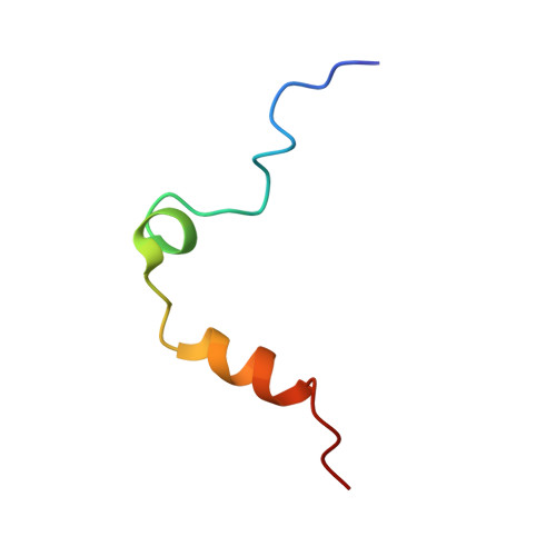
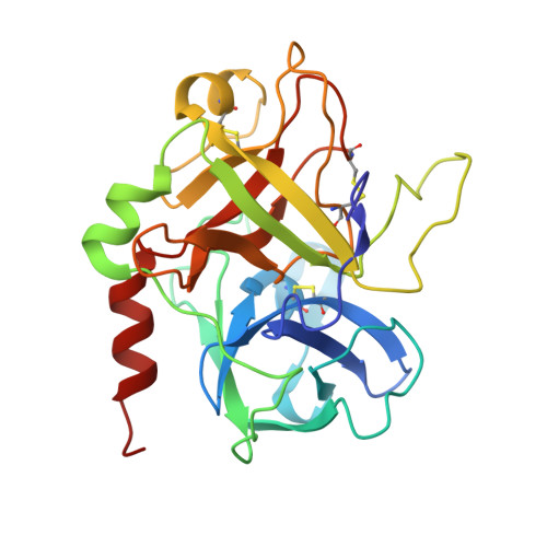
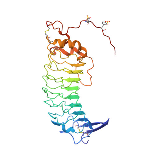
Modulation of alpha-thrombin function by distinct interactions with platelet glycoprotein Ibalpha

Celikel, R.McClintock, R.A.Roberts, J.R.Mendolicchio, G.L.Ware, J.Varughese, K.I.Ruggeri, Z.M.

(2003) Science 301: 218-221

  • DOI: https://doi.org/10.1126/science.1084183
  • Primary Citation of Related Structures:  
    1OOK, 1P9A

  • PubMed Abstract: 

    Thrombin bound to platelets contributes to stop bleeding and, in pathological conditions, may cause vascular thrombosis. We have determined the structure of platelet glycoprotein Ibalpha (GpIbalpha) bound to thrombin at 2.3 angstrom resolution and defined two sites in GpIbalpha that bind to exosite II and exosite I of two distinct alpha-thrombin molecules, respectively. GpIbalpha occupancy may be sequential, as the site binding to alpha-thrombin exosite I appears to be cryptic in the unoccupied receptor but exposed when a first thrombin molecule is bound through exosite II. These interactions may modulate alpha-thrombin function by mediating GpIbalpha clustering and cleavage of protease-activated receptors, which promote platelet activation, while limiting fibrinogen clotting through blockade of exosite I.


  • Organizational Affiliation

    Roon Research Center for Arteriosclerosis and Thrombosis, Division of Experimental Thrombosis and Hemostasis, Scripps Research Institute, 10550 North Torrey Pines Road, La Jolla, CA 92037, USA.


Macromolecules
Find similar proteins by:  (by identity cutoff)  |  3D Structure
Entity ID: 1
MoleculeChains Sequence LengthOrganismDetailsImage
Human Alpha Thrombin36Homo sapiensMutation(s): 0 
EC: 3.4.21.5
UniProt & NIH Common Fund Data Resources
Find proteins for P00734 (Homo sapiens)
Explore P00734 
Go to UniProtKB:  P00734
PHAROS:  P00734
GTEx:  ENSG00000180210 
Entity Groups  
Sequence Clusters30% Identity50% Identity70% Identity90% Identity95% Identity100% Identity
UniProt GroupP00734
Sequence Annotations
Expand
  • Reference Sequence
Find similar proteins by:  (by identity cutoff)  |  3D Structure
Entity ID: 2
MoleculeChains Sequence LengthOrganismDetailsImage
Human Alpha Thrombin259Homo sapiensMutation(s): 0 
EC: 3.4.21.5
UniProt & NIH Common Fund Data Resources
Find proteins for P00734 (Homo sapiens)
Explore P00734 
Go to UniProtKB:  P00734
PHAROS:  P00734
GTEx:  ENSG00000180210 
Entity Groups  
Sequence Clusters30% Identity50% Identity70% Identity90% Identity95% Identity100% Identity
UniProt GroupP00734
Sequence Annotations
Expand
  • Reference Sequence

Find similar proteins by:  Sequence   |   3D Structure  

Entity ID: 3
MoleculeChains Sequence LengthOrganismDetailsImage
PHE-PRO-ARG-ChloromethylketoneC [auth P]3N/AMutation(s): 0 
Sequence Annotations
Expand
  • Reference Sequence
Find similar proteins by:  (by identity cutoff)  |  3D Structure
Entity ID: 4
MoleculeChains Sequence LengthOrganismDetailsImage
Platelet glycoprotein Ib alpha chain precursorD [auth G]290Homo sapiensMutation(s): 3 
Gene Names: GP1BA
UniProt & NIH Common Fund Data Resources
Find proteins for P07359 (Homo sapiens)
Explore P07359 
Go to UniProtKB:  P07359
PHAROS:  P07359
GTEx:  ENSG00000185245 
Entity Groups  
Sequence Clusters30% Identity50% Identity70% Identity90% Identity95% Identity100% Identity
UniProt GroupP07359
Sequence Annotations
Expand
  • Reference Sequence
Oligosaccharides

Help

Entity ID: 5
MoleculeChains Length2D Diagram Glycosylation3D Interactions
alpha-D-mannopyranose-(1-3)-[alpha-D-mannopyranose-(1-6)]alpha-D-mannopyranose-(1-4)-2-acetamido-2-deoxy-beta-D-glucopyranose-(1-4)-2-acetamido-2-deoxy-beta-D-glucopyranoseE [auth C]5N-Glycosylation
Glycosylation Resources
GlyTouCan:  G21381MC
GlyCosmos:  G21381MC
GlyGen:  G21381MC
Small Molecules
Ligands 2 Unique
IDChains Name / Formula / InChI Key2D Diagram3D Interactions
NAG
Query on NAG

Download Ideal Coordinates CCD File 
G
2-acetamido-2-deoxy-beta-D-glucopyranose
C8 H15 N O6
OVRNDRQMDRJTHS-FMDGEEDCSA-N
CL
Query on CL

Download Ideal Coordinates CCD File 
F [auth B]CHLORIDE ION
Cl
VEXZGXHMUGYJMC-UHFFFAOYSA-M
Modified Residues  1 Unique
IDChains TypeFormula2D DiagramParent
TYS
Query on TYS
D [auth G]L-PEPTIDE LINKINGC9 H11 N O6 STYR
Experimental Data & Validation

Experimental Data

  • Method: X-RAY DIFFRACTION
  • Resolution: 2.30 Å
  • R-Value Free: 0.260 
  • R-Value Work: 0.212 
  • R-Value Observed: 0.214 
  • Space Group: P 43 21 2
Unit Cell:
Length ( Å )Angle ( ˚ )
a = 67.649α = 90
b = 67.649β = 90
c = 328.578γ = 90
Software Package:
Software NamePurpose
HKL-2000data collection
SCALEPACKdata scaling
CNSrefinement
HKL-2000data reduction
CNSphasing

Structure Validation

View Full Validation Report


Ligand Structure Quality Assessment 


Entry History 

Deposition Data

Revision History  (Full details and data files)

  • Version 1.0: 2003-07-22
    Type: Initial release
  • Version 1.1: 2008-04-29
    Changes: Version format compliance
  • Version 1.2: 2011-07-13
    Changes: Non-polymer description, Version format compliance
  • Version 2.0: 2020-07-29
    Type: Remediation
    Reason: Carbohydrate remediation
    Changes: Atomic model, Data collection, Database references, Derived calculations, Structure summary
  • Version 2.1: 2021-10-27
    Changes: Database references, Structure summary