High-resolution structure of the oligomerization domain of p53 by multidimensional NMR.
Clore, G.M., Omichinski, J.G., Sakaguchi, K., Zambrano, N., Sakamoto, H., Appella, E., Gronenborn, A.M.(1994) Science 265: 386-391
- PubMed: 8023159
- DOI: https://doi.org/10.1126/science.8023159
- Primary Citation of Related Structures:
1OLG, 1OLH - PubMed Abstract:
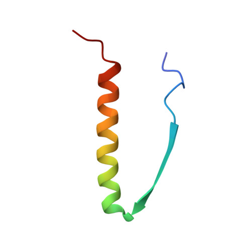
The three-dimensional structure of the oligomerization domain (residues 319 to 360) of the tumor suppressor p53 has been solved by multidimensional heteronuclear magnetic resonance (NMR) spectroscopy. The domain forms a 20-kilodalton symmetric tetramer with a topology made up from a dimer of dimers. The two primary dimers each comprise two antiparallel helices linked by an antiparallel beta sheet. One beta strand and one helix are contributed from each monomer. The interface between the two dimers forming the tetramer is mediated solely by helix-helix contacts. The overall result is a symmetric, four-helix bundle with adjacent helices oriented antiparallel to each other and with the two antiparallel beta sheets located on opposing faces of the molecule. The tetramer is stabilized not only by hydrophobic interactions within the protein core but also by a number of electrostatic interactions. The implications of the structure of the tetramer for the biological function of p53 are discussed.
Organizational Affiliation:
Laboratory of Chemical Physics, National Institute of Diabetes and Digestive and Kidney Diseases, National Institutes of Health, Bethesda, MD 20892.