1NWM

GAT domain of human GGA1


Experimental Data Snapshot

  • Method: X-RAY DIFFRACTION
  • Resolution: 2.40 Å
  • R-Value Free: 0.286 
  • R-Value Work: 0.252 
  • R-Value Observed: 0.254 

wwPDB Validation   3D Report Full Report


This is version 1.3 of the entry. See complete history


Literature

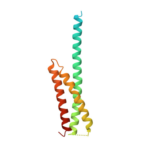
Structure of the GAT domain of human GGA1: a syntaxin amino-terminal domain fold in an endosomal trafficking adaptor.

Suer, S.Misra, S.Saidi, L.F.Hurley, J.H.

(2003) Proc Natl Acad Sci U S A 100: 4451-4456

  • DOI: https://doi.org/10.1073/pnas.0831133100
  • Primary Citation of Related Structures:  
    1NWM

  • PubMed Abstract: 

    The Golgi-associated, gamma-adaptin homologous, ADP-ribosylation factor (ARF)-interacting proteins (GGAs) are adaptors that sort receptors from the trans-Golgi network into the endosomallysosomal pathway. The GGAs and TOM1 (GAT) domains of the GGAs are responsible for their ARF-dependent localization. The 2.4-A crystal structure of the GAT domain of human GGA1 reveals a three-helix bundle, with a long N-terminal helical extension that is not conserved in GAT domains that do not bind ARF. The ARF binding site is located in the N-terminal extension and is separate from the core three-helix bundle. An unanticipated structural similarity to the N-terminal domain of syntaxin 1a was discovered, comprising the entire three-helix bundle. A conserved binding site on helices 2 and 3 of the GAT domain three-helix bundle is predicted to interact with coiled-coil-containing proteins. We propose that the GAT domain is descended from the same ancestor as the syntaxin 1a N-terminal domain, and that both protein families share a common function in binding coiled-coil domain proteins.


  • Organizational Affiliation

    Laboratory of Molecular Biology, National Institute of Diabetes and Digestive and Kidney Diseases, National Institutes of Health, Department of Health and Human Services, Bethesda, MD 20892, USA.


Macromolecules
Find similar proteins by:  (by identity cutoff)  |  3D Structure
Entity ID: 1
MoleculeChains Sequence LengthOrganismDetailsImage
ADP-ribosylation factor binding protein GGA1A [auth X]142Homo sapiensMutation(s): 0 
UniProt & NIH Common Fund Data Resources
Find proteins for Q9UJY5 (Homo sapiens)
Explore Q9UJY5 
Go to UniProtKB:  Q9UJY5
PHAROS:  Q9UJY5
GTEx:  ENSG00000100083 
Entity Groups  
Sequence Clusters30% Identity50% Identity70% Identity90% Identity95% Identity100% Identity
UniProt GroupQ9UJY5
Sequence Annotations
Expand
  • Reference Sequence
Experimental Data & Validation

Experimental Data

  • Method: X-RAY DIFFRACTION
  • Resolution: 2.40 Å
  • R-Value Free: 0.286 
  • R-Value Work: 0.252 
  • R-Value Observed: 0.254 
  • Space Group: H 3
Unit Cell:
Length ( Å )Angle ( ˚ )
a = 76.66α = 90
b = 76.66β = 90
c = 60.816γ = 120
Software Package:
Software NamePurpose
REFMACrefinement
HKL-2000data reduction
SCALEPACKdata scaling
CNSphasing

Structure Validation

View Full Validation Report


Entry History 

Deposition Data

Revision History  (Full details and data files)

  • Version 1.0: 2003-03-25
    Type: Initial release
  • Version 1.1: 2008-04-29
    Changes: Version format compliance
  • Version 1.2: 2011-07-13
    Changes: Version format compliance
  • Version 1.3: 2024-02-14
    Changes: Data collection, Database references