1NO9

Design of weakly basic thrombin inhibitors incorporating novel P1 binding functions: molecular and X-ray crystallographic studies.


Experimental Data Snapshot

  • Method: X-RAY DIFFRACTION
  • Resolution: 1.90 Å
  • R-Value Free: 0.204 
  • R-Value Work: 0.183 

wwPDB Validation   3D Report Full Report


Ligand Structure Quality Assessment 


This is version 1.5 of the entry. See complete history


Literature

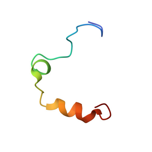
DESIGN OF WEAKLY BASIC THROMBIN INHIBITORS INCORPORATING NOVEL P1 BINDING FUNCTIONS: MOLECULAR AND X-RAY CRYSTALLOGRAPHIC STUDIES

De Simone, G.Menchise, V.Omaggio, S.Pedone, C.Scozzafava, A.Supuran, C.T.

(2003) Biochemistry 42: 9013-9021

  • DOI: https://doi.org/10.1021/bi020512l
  • Primary Citation of Related Structures:  
    1NO9

  • PubMed Abstract: 

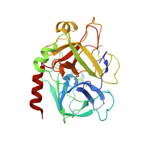
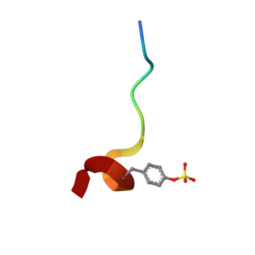
    To prepare weakly basic thrombin inhibitors with modified S1 anchoring groups, two series of compounds were synthesized by reaction of guanidine or aminoguanidine with acyl halides and N,N-disubstituted carbamoyl chlorides. pK(a) measurements of these acylated guanidines/aminoguanidines showed a reduced basicity, with pK(a) values in the range of 8.4-8.7. These molecules typically showed inhibition constants in the range of 150-425 nM against thrombin and 360-965 nM against trypsin, even though some bulky derivatives, such as N,N-diphenylcarbamoylguanidine/aminoguanidine and their congeners, showed much stronger thrombin inhibitory activity, with inhibition constants in the range of 24-42 nM. Unexpectedly, very long incubation times with both proteases revealed that aminoguanidine derivatives behaved as irreversible inhibitors. To assess the molecular basis responsible for the high affinity observed for these molecules toward thrombin, the crystal structure of the thrombin-hirugen-N,N-diphenylcarbamoylaminoguanidine complex has been solved at 1.90 A resolution. The structural analysis of the complex revealed an unexpected interaction mode with the protease, resulting in an N,N-diphenylcarbamoyl intermediate covalently bound to the catalytic serine as a consequence of its hydrolysis together with the release of the aminoguanidine moiety. Surprisingly, in this covalent adduct a phenyl group was found in the S1 specificity pocket, which usually recognizes positively charged residues. These findings provide new insights in the design of low basicity serine protease inhibitors.


  • Organizational Affiliation

    Laboratorio di Chimica Inorganica e Bioinorganica, Università degli Studi, via della Lastruccia 3, Rm 188, Polo Scientifico, 50019 Sesto Fiorentino, Florence, Italy. gmg@chemistry.unina.it


Macromolecules
Find similar proteins by:  (by identity cutoff)  |  3D Structure
Entity ID: 1
MoleculeChains Sequence LengthOrganismDetailsImage
Alpha ThrombinA [auth L]36Homo sapiensMutation(s): 0 
EC: 3.4.21.5
UniProt & NIH Common Fund Data Resources
Find proteins for P00734 (Homo sapiens)
Explore P00734 
Go to UniProtKB:  P00734
PHAROS:  P00734
GTEx:  ENSG00000180210 
Entity Groups  
Sequence Clusters30% Identity50% Identity70% Identity90% Identity95% Identity100% Identity
UniProt GroupP00734
Sequence Annotations
Expand
  • Reference Sequence
Find similar proteins by:  (by identity cutoff)  |  3D Structure
Entity ID: 2
MoleculeChains Sequence LengthOrganismDetailsImage
Alpha ThrombinB [auth H]259Homo sapiensMutation(s): 0 
EC: 3.4.21.5
UniProt & NIH Common Fund Data Resources
Find proteins for P00734 (Homo sapiens)
Explore P00734 
Go to UniProtKB:  P00734
PHAROS:  P00734
GTEx:  ENSG00000180210 
Entity Groups  
Sequence Clusters30% Identity50% Identity70% Identity90% Identity95% Identity100% Identity
UniProt GroupP00734
Sequence Annotations
Expand
  • Reference Sequence

Find similar proteins by:  Sequence   |   3D Structure  

Entity ID: 3
MoleculeChains Sequence LengthOrganismDetailsImage
hirugenC [auth I]11Hirudo medicinalisMutation(s): 0 
UniProt
Find proteins for P01050 (Hirudo medicinalis)
Explore P01050 
Go to UniProtKB:  P01050
Entity Groups  
Sequence Clusters30% Identity50% Identity70% Identity90% Identity95% Identity100% Identity
UniProt GroupP01050
Sequence Annotations
Expand
  • Reference Sequence
Small Molecules
Ligands 2 Unique
IDChains Name / Formula / InChI Key2D Diagram3D Interactions
4ND
Query on 4ND

Download Ideal Coordinates CCD File 
E [auth H]N4-(N,N-DIPHENYLCARBAMOYL)-AMINOGUANIDINE
C14 H15 N5 O
QLXOHIUDKNRVBZ-UHFFFAOYSA-N
NAG
Query on NAG

Download Ideal Coordinates CCD File 
D [auth H]2-acetamido-2-deoxy-beta-D-glucopyranose
C8 H15 N O6
OVRNDRQMDRJTHS-FMDGEEDCSA-N
Modified Residues  1 Unique
IDChains TypeFormula2D DiagramParent
TYS
Query on TYS
C [auth I]L-PEPTIDE LINKINGC9 H11 N O6 STYR
Binding Affinity Annotations 
IDSourceBinding Affinity
4ND PDBBind:  1NO9 Ki: 24 (nM) from 1 assay(s)
Experimental Data & Validation

Experimental Data

  • Method: X-RAY DIFFRACTION
  • Resolution: 1.90 Å
  • R-Value Free: 0.204 
  • R-Value Work: 0.183 
  • Space Group: C 1 2 1
Unit Cell:
Length ( Å )Angle ( ˚ )
a = 71.413α = 90
b = 72.192β = 100.57
c = 72.705γ = 90
Software Package:
Software NamePurpose
DENZOdata reduction
SCALEPACKdata scaling
CNSrefinement
CNSphasing

Structure Validation

View Full Validation Report


Ligand Structure Quality Assessment 


Entry History 

Deposition Data

Revision History  (Full details and data files)

  • Version 1.0: 2003-08-26
    Type: Initial release
  • Version 1.1: 2008-04-29
    Changes: Version format compliance
  • Version 1.2: 2011-07-13
    Changes: Atomic model, Database references, Derived calculations, Non-polymer description, Structure summary, Version format compliance
  • Version 1.3: 2020-07-29
    Type: Remediation
    Reason: Carbohydrate remediation
    Changes: Advisory, Data collection, Derived calculations, Structure summary
  • Version 1.4: 2023-08-16
    Changes: Advisory, Data collection, Database references, Refinement description, Structure summary
  • Version 1.5: 2023-11-15
    Changes: Data collection