1NKF

CALCIUM-BINDING PEPTIDE, NMR, 30 STRUCTURES


Experimental Data Snapshot

  • Method: SOLUTION NMR
  • Conformers Calculated: 100 
  • Conformers Submitted: 30 
  • Selection Criteria: ENERGY, AGREEMENT WITH EXPERIMENTAL DATA 

wwPDB Validation   3D Report Full Report


This is version 1.3 of the entry. See complete history


Literature

Alpha-helix nucleation by a calcium-binding peptide loop.

Siedlecka, M.Goch, G.Ejchart, A.Sticht, H.Bierzyski, A.

(1999) Proc Natl Acad Sci U S A 96: 903-908

  • DOI: https://doi.org/10.1073/pnas.96.3.903
  • Primary Citation of Related Structures:  
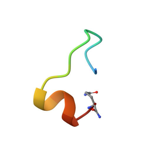
    1NKF

  • PubMed Abstract: 

    A 12-residue peptide AcDKDGDGYISAAENH2 analogous to the third calcium-binding loop of calmodulin strongly coordinates lanthanide ions (K = 10(5) M-1). When metal saturated, the peptide adopts a very rigid structure, the same as in the native protein, with three last residues AAE fixed in the alpha-helical conformation. Therefore, the peptide provides an ideal helix nucleation site for peptide segments attached to its C terminus. NMR and CD investigations of peptide AcDKDGDGYISAAEAAAQNH2 presented in this paper show that residues A13-Q16 form an alpha-helix of very high stability when the La3+ ion is bound to the D1-E12 loop. In fact, the lowest estimates of the helix content in this segment give values of at least 80% at 1 degreesC and 70% at 25 degreesC. This finding is not compatible with existing helix-coil transition theories and helix propagation parameters, s, reported in the literature. We conclude, therefore, that the initial steps of helix propagation are characterized by much larger s values, whereas helix nucleation is even more unfavorable than is believed. In light of our findings, thermodynamics of the nascent alpha-helices is discussed. The problem of CD spectra of very short alpha-helices is also addressed.


  • Organizational Affiliation

    Institute of Biochemistry and Biophysics, Polish Academy of Sciences, 02-106 Warszawa, ul. Pawińskiego 5A, Poland.


Macromolecules

Find similar proteins by:  Sequence   |   3D Structure  

Entity ID: 1
MoleculeChains Sequence LengthOrganismDetailsImage
CALCIUM-BINDING HEXADECAPEPTIDE18Homo sapiensMutation(s): 0 
UniProt & NIH Common Fund Data Resources
Find proteins for P0DP23 (Homo sapiens)
Explore P0DP23 
Go to UniProtKB:  P0DP23
PHAROS:  P0DP23
GTEx:  ENSG00000198668 
Entity Groups  
Sequence Clusters30% Identity50% Identity70% Identity90% Identity95% Identity100% Identity
UniProt GroupP0DP23
Sequence Annotations
Expand
  • Reference Sequence
Small Molecules
Ligands 1 Unique
IDChains Name / Formula / InChI Key2D Diagram3D Interactions
LA
Query on LA

Download Ideal Coordinates CCD File 
B [auth A]LANTHANUM (III) ION
La
CZMAIROVPAYCMU-UHFFFAOYSA-N
Experimental Data & Validation

Experimental Data

  • Method: SOLUTION NMR
  • Conformers Calculated: 100 
  • Conformers Submitted: 30 
  • Selection Criteria: ENERGY, AGREEMENT WITH EXPERIMENTAL DATA 

Structure Validation

View Full Validation Report


Entry History 

Deposition Data

Revision History  (Full details and data files)

  • Version 1.0: 1999-02-16
    Type: Initial release
  • Version 1.1: 2008-03-24
    Changes: Version format compliance
  • Version 1.2: 2011-07-13
    Changes: Version format compliance
  • Version 1.3: 2022-02-23
    Changes: Database references, Derived calculations, Other