1NAF

Crystal structure of the human GGA1 GAT domain


Experimental Data Snapshot

  • Method: X-RAY DIFFRACTION
  • Resolution: 2.80 Å
  • R-Value Free: 0.283 
  • R-Value Work: 0.248 
  • R-Value Observed: 0.248 

wwPDB Validation   3D Report Full Report


This is version 1.3 of the entry. See complete history


Literature

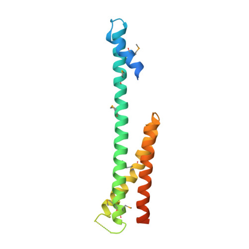
The Structure of the GGA1-GAT Domain Reveals the Molecular Basis for ARF Binding and Membrane Association of GGAs

Collins, B.M.Watson, P.J.Owen, D.J.

(2003) Dev Cell 4: 321-332

  • DOI: https://doi.org/10.1016/s1534-5807(03)00037-6
  • Primary Citation of Related Structures:  
    1NAF

  • PubMed Abstract: 

    The GGAs are a family of clathrin adaptor proteins involved in vesicular transport between the trans-Golgi network and endosomal system. Here we confirm reports that GGAs are targeted to the Golgi via interaction between the GGA-GAT domain and ARF-GTP, and we present the structure of the GAT domain of human GGA1, completing the structural description of the folded domains of GGA proteins. The GGA-GAT domain possesses an all alpha-helical fold with a "paper clip" topology comprising two independent subdomains. Structure-based mutagenesis demonstrates that ARF1-GTP binding by GGAs is exclusively governed by the N-terminal "hook" subdomain, and, using an in vitro recruitment assay, we show that ARF-GTP binding by this small structure is required and sufficient for Golgi targeting of GGAs.


  • Organizational Affiliation

    Department of Clinical Biochemistry, University of Cambridge, Hills Road, CB2 2XY, Cambridge, United Kingdom. bmc25@cam.ac.uk


Macromolecules
Find similar proteins by:  (by identity cutoff)  |  3D Structure
Entity ID: 1
MoleculeChains Sequence LengthOrganismDetailsImage
ADP-ribosylation factor binding protein GGA1158Homo sapiensMutation(s): 0 
UniProt & NIH Common Fund Data Resources
Find proteins for Q9UJY5 (Homo sapiens)
Explore Q9UJY5 
Go to UniProtKB:  Q9UJY5
PHAROS:  Q9UJY5
GTEx:  ENSG00000100083 
Entity Groups  
Sequence Clusters30% Identity50% Identity70% Identity90% Identity95% Identity100% Identity
UniProt GroupQ9UJY5
Sequence Annotations
Expand
  • Reference Sequence
Small Molecules
Modified Residues  1 Unique
IDChains TypeFormula2D DiagramParent
MSE
Query on MSE
A
L-PEPTIDE LINKINGC5 H11 N O2 SeMET
Experimental Data & Validation

Experimental Data

  • Method: X-RAY DIFFRACTION
  • Resolution: 2.80 Å
  • R-Value Free: 0.283 
  • R-Value Work: 0.248 
  • R-Value Observed: 0.248 
  • Space Group: P 63
Unit Cell:
Length ( Å )Angle ( ˚ )
a = 82.7α = 90
b = 82.7β = 90
c = 69.9γ = 120
Software Package:
Software NamePurpose
MOSFLMdata reduction
SCALAdata scaling
SHARPphasing
CNSrefinement
CCP4data scaling

Structure Validation

View Full Validation Report


Entry History 

Deposition Data

Revision History  (Full details and data files)

  • Version 1.0: 2003-03-25
    Type: Initial release
  • Version 1.1: 2008-04-28
    Changes: Version format compliance
  • Version 1.2: 2011-07-13
    Changes: Version format compliance
  • Version 1.3: 2018-02-14
    Changes: Experimental preparation, Structure summary