Three-dimensional domain swapping in the folded and molten-globule states of cystatins, an amyloid-forming structural superfamily
Staniforth, R.A., Giannini, S., Higgins, L.D., Conroy, M.J., Hounslow, A.M., Jerala, R., Craven, C.J., Waltho, J.P.(2001) EMBO J 20: 4774-4781
- PubMed: 11532941
- DOI: https://doi.org/10.1093/emboj/20.17.4774
- Primary Citation of Related Structures:
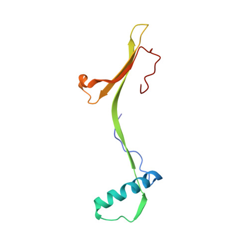
1N9J - PubMed Abstract:
Cystatins, an amyloid-forming structural superfamily, form highly stable, domain-swapped dimers at physiological protein concentrations. In chicken cystatin, the active monomer is a kinetic trap en route to dimerization, and any changes in solution conditions or mutations that destabilize the folded state shorten the lifetime of the monomeric form. In such circumstances, amyloidogenesis will start from conditions where a domain-swapped dimer is the most prevalent species. Domain swapping occurs by a rearrangement of loop I, generating the new intermonomer interface between strands 2 and 3. The transition state for dimerization has a high level of hydrophobic group exposure, indicating that gross conformational perturbation is required for domain swapping to occur. Dimerization also occurs when chicken cystatin is in its reduced, molten-globule state, implying that the organization of secondary structure in this state mirrors that in the folded state and that domain swapping is not limited to the folded states of proteins. Although the interface between cystatin-fold units is poorly defined for cystatin A, the dimers are the appropriate size to account for the electron-dense regions in amyloid protofilaments.
Organizational Affiliation:
Krebs Institute, Department of Molecular Biology and Biotechnology, University of Sheffield, Transgenomic Ltd, Western Bank, Sheffield S10 2TN, UK. r.a.staniforth@shef.ac.uk