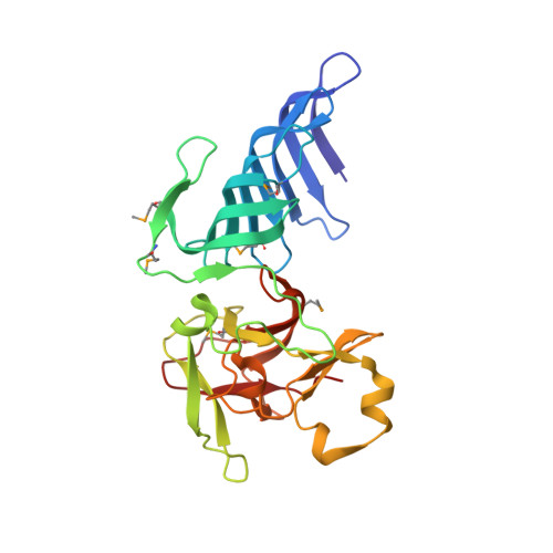
1MUF

Structure of histone H3 K4-specific methyltransferase SET7/9


Experimental Data Snapshot

  • Method: X-RAY DIFFRACTION
  • Resolution: 2.26 Å
  • R-Value Free: 0.249 
  • R-Value Work: 0.229 
  • R-Value Observed: 0.229 

wwPDB Validation   3D Report Full Report


This is version 1.3 of the entry. See complete history


Literature

The active site of the SET domain is constructed on a knot

Jacobs, S.A.Harp, J.M.Devarakonda, S.Kim, Y.Rastinejad, F.Khorasanizadeh, S.

(2002) Nat Struct Biol 9: 833-838

  • DOI: https://doi.org/10.1038/nsb861
  • Primary Citation of Related Structures:  
    1MT6, 1MUF

  • PubMed Abstract: 

    The SET domain contains the catalytic center of lysine methyltransferases that target the N-terminal tails of histones and regulate chromatin function. Here we report the structure of the SET7/9 protein in the absence and presence of its cofactor product, S-adenosyl-L-homocysteine (AdoHcy). A knot within the SET domain helps form the methyltransferase active site, where AdoHcy binds and lysine methylation is likely to occur. A structure-guided comparison of sequences within the SET protein family suggests that the knot substructure and active site environment are conserved features of the SET domain.


  • Organizational Affiliation

    Department of Biochemistry and Molecular Genetics, University of Virginia Health System, Charlottesville, Virginia 22908, USA.


Macromolecules
Find similar proteins by:  (by identity cutoff)  |  3D Structure
Entity ID: 1
MoleculeChains Sequence LengthOrganismDetailsImage
SET9257Homo sapiensMutation(s): 6 
EC: 2.1.1.43
UniProt & NIH Common Fund Data Resources
Find proteins for Q8WTS6 (Homo sapiens)
Explore Q8WTS6 
Go to UniProtKB:  Q8WTS6
PHAROS:  Q8WTS6
GTEx:  ENSG00000145391 
Entity Groups  
Sequence Clusters30% Identity50% Identity70% Identity90% Identity95% Identity100% Identity
UniProt GroupQ8WTS6
Sequence Annotations
Expand
  • Reference Sequence
Small Molecules
Modified Residues  1 Unique
IDChains TypeFormula2D DiagramParent
MSE
Query on MSE
A
L-PEPTIDE LINKINGC5 H11 N O2 SeMET
Experimental Data & Validation

Experimental Data

  • Method: X-RAY DIFFRACTION
  • Resolution: 2.26 Å
  • R-Value Free: 0.249 
  • R-Value Work: 0.229 
  • R-Value Observed: 0.229 
  • Space Group: P 64 2 2
Unit Cell:
Length ( Å )Angle ( ˚ )
a = 110.973α = 90
b = 110.973β = 90
c = 96.05γ = 120
Software Package:
Software NamePurpose
HKL-2000data collection
SCALEPACKdata scaling
SOLVEphasing
RESOLVEmodel building
CNSrefinement
HKL-2000data reduction
RESOLVEphasing

Structure Validation

View Full Validation Report


Entry History 

Deposition Data

Revision History  (Full details and data files)

  • Version 1.0: 2002-11-06
    Type: Initial release
  • Version 1.1: 2008-04-28
    Changes: Version format compliance
  • Version 1.2: 2011-07-13
    Changes: Version format compliance
  • Version 1.3: 2011-11-16
    Changes: Atomic model