1MB8

Crystal Structure of the actin binding domain of plectin


Experimental Data Snapshot

  • Method: X-RAY DIFFRACTION
  • Resolution: 2.15 Å
  • R-Value Free: 0.261 
  • R-Value Work: 0.212 

wwPDB Validation   3D Report Full Report


This is version 1.3 of the entry. See complete history


Literature

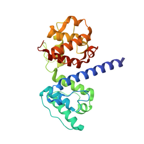
Structural and Functional Analysis of the Actin Binding Domain of Plectin Suggests Alternative Mechanisms for Binding to F-Actin and Integrin Beta4

Garcia-Alvarez, B.Bobkov, A.Sonnenberg, A.de Pereda, J.M.

(2003) Structure 11: 615-625

  • DOI: https://doi.org/10.1016/s0969-2126(03)00090-x
  • Primary Citation of Related Structures:  
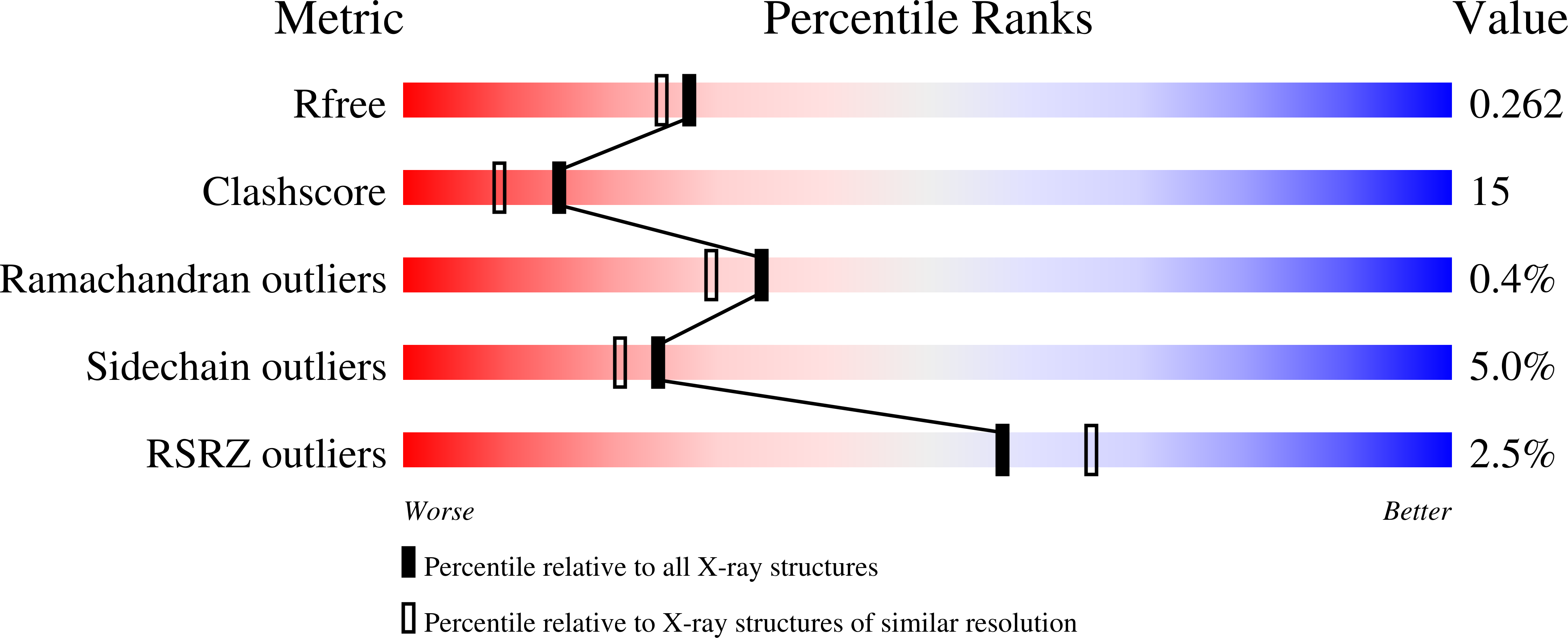
    1MB8

  • PubMed Abstract: 

    Plectin is a widely expressed cytoskeletal linker. Here we report the crystal structure of the actin binding domain of plectin and show that this region is sufficient for interaction with F-actin or the cytoplasmic region of integrin alpha6beta4. The structure is formed by two calponin homology domains arranged in a closed conformation. We show that binding to F-actin induces a conformational change in plectin that is inhibited by an engineered interdomain disulfide bridge. A two-step induced fit mechanism involving binding and subsequent domain rearrangement is proposed. In contrast, interaction with integrin alpha6beta4 occurs in a closed conformation. Competitive binding of plectin to F-actin and integrin alpha6beta4 may rely on the observed alternative binding mechanisms and involve both allosteric and steric factors.


  • Organizational Affiliation

    Program on Cell Adhesion, The Burnham Institute, 10901 North Torrey Pines Road, La Jolla, CA 92037, USA.


Macromolecules
Find similar proteins by:  (by identity cutoff)  |  3D Structure
Entity ID: 1
MoleculeChains Sequence LengthOrganismDetailsImage
Plectin243Homo sapiensMutation(s): 0 
UniProt & NIH Common Fund Data Resources
Find proteins for Q15149 (Homo sapiens)
Explore Q15149 
Go to UniProtKB:  Q15149
PHAROS:  Q15149
GTEx:  ENSG00000178209 
Entity Groups  
Sequence Clusters30% Identity50% Identity70% Identity90% Identity95% Identity100% Identity
UniProt GroupQ15149
Sequence Annotations
Expand
  • Reference Sequence
Experimental Data & Validation

Experimental Data

  • Method: X-RAY DIFFRACTION
  • Resolution: 2.15 Å
  • R-Value Free: 0.261 
  • R-Value Work: 0.212 
  • Space Group: P 21 21 21
Unit Cell:
Length ( Å )Angle ( ˚ )
a = 33.597α = 90
b = 79.283β = 90
c = 82.37γ = 90
Software Package:
Software NamePurpose
DENZOdata reduction
SCALEPACKdata scaling
CNSrefinement
CNSphasing

Structure Validation

View Full Validation Report


Entry History 

Deposition Data

Revision History  (Full details and data files)

  • Version 1.0: 2003-06-10
    Type: Initial release
  • Version 1.1: 2008-04-28
    Changes: Version format compliance
  • Version 1.2: 2011-07-13
    Changes: Version format compliance
  • Version 1.3: 2024-02-14
    Changes: Data collection, Database references