1KSW

Structure of Human c-Src Tyrosine Kinase (Thr338Gly Mutant) in Complex with N6-benzyl ADP


Experimental Data Snapshot

  • Method: X-RAY DIFFRACTION
  • Resolution: 2.80 Å
  • R-Value Free: 0.294 
  • R-Value Work: 0.231 

wwPDB Validation   3D Report Full Report


Ligand Structure Quality Assessment 


This is version 1.5 of the entry. See complete history


Literature

Mutant tyrosine kinases with unnatural nucleotide specificity retain the structure and phospho-acceptor specificity of the wild-type enzyme.

Witucki, L.A.Huang, X.Shah, K.Liu, Y.Kyin, S.Eck, M.J.Shokat, K.M.

(2002) Chem Biol 9: 25-33

  • DOI: https://doi.org/10.1016/s1074-5521(02)00091-1
  • Primary Citation of Related Structures:  
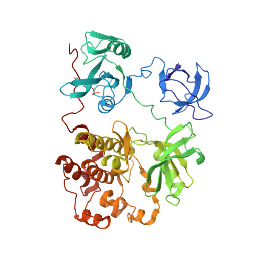
    1KSW

  • PubMed Abstract: 

    The direct substrates of one protein kinase in a cell can be identified by mutation of the ATP binding pocket to allow an unnatural ATP analog to be accepted exclusively by the engineered kinase. Here, we present structural and functional assessment of peptide specificity of mutant protein kinases with unnatural ATP analogs. The crystal structure (2.8 A resolution) of c-Src (T338G) with N(6)-(benzyl) ADP bound shows that the creation of a unique nucleotide binding pocket does not alter the phospho-acceptor binding site of the kinase. A panel of optimal peptide substrates of defined sequence, as well as a degenerate peptide library, was utilized to assess the phospho-acceptor specificity of the engineered "traceable" kinases. The specificity profiles for the mutant kinases were found to be identical to those of their wild-type counterparts.


  • Organizational Affiliation

    Department of Cellular and Molecular Pharmacology, Box 0450, University of California, San Francisco 94143-0450, USA.


Macromolecules
Find similar proteins by:  (by identity cutoff)  |  3D Structure
Entity ID: 1
MoleculeChains Sequence LengthOrganismDetailsImage
PROTO-ONCOGENE TYROSINE-PROTEIN KINASE SRC452Homo sapiensMutation(s): 2 
EC: 2.7.1.112
UniProt & NIH Common Fund Data Resources
Find proteins for P12931 (Homo sapiens)
Explore P12931 
Go to UniProtKB:  P12931
PHAROS:  P12931
GTEx:  ENSG00000197122 
Entity Groups  
Sequence Clusters30% Identity50% Identity70% Identity90% Identity95% Identity100% Identity
UniProt GroupP12931
Sequence Annotations
Expand
  • Reference Sequence
Small Molecules
Ligands 1 Unique
IDChains Name / Formula / InChI Key2D Diagram3D Interactions
NBS
Query on NBS

Download Ideal Coordinates CCD File 
B [auth A]N6-BENZYL ADENOSINE-5'-DIPHOSPHATE
C17 H21 N5 O10 P2
MRHGMAGSDAQUFH-LSCFUAHRSA-N
Modified Residues  1 Unique
IDChains TypeFormula2D DiagramParent
PTR
Query on PTR
A
L-PEPTIDE LINKINGC9 H12 N O6 PTYR
Binding Affinity Annotations 
IDSourceBinding Affinity
PTR BindingDB:  1KSW -TΔS: -1.81e+1 (kJ/mol) from 1 assay(s)
ΔG: -2.14e+1 (kJ/mol) from 1 assay(s)
Experimental Data & Validation

Experimental Data

  • Method: X-RAY DIFFRACTION
  • Resolution: 2.80 Å
  • R-Value Free: 0.294 
  • R-Value Work: 0.231 
  • Space Group: P 21 21 21
Unit Cell:
Length ( Å )Angle ( ˚ )
a = 50.81α = 90
b = 87.68β = 90
c = 106.28γ = 90
Software Package:
Software NamePurpose
AMoREphasing
X-PLORrefinement
DENZOdata reduction
SCALEPACKdata scaling

Structure Validation

View Full Validation Report


Ligand Structure Quality Assessment 


Entry History 

Deposition Data

Revision History  (Full details and data files)

  • Version 1.0: 2002-02-27
    Type: Initial release
  • Version 1.1: 2008-04-27
    Changes: Version format compliance
  • Version 1.2: 2011-07-13
    Changes: Version format compliance
  • Version 1.3: 2021-10-27
    Changes: Database references, Derived calculations
  • Version 1.4: 2023-08-16
    Changes: Data collection, Refinement description
  • Version 1.5: 2023-11-15
    Changes: Data collection