1JMT

X-ray Structure of a Core U2AF65/U2AF35 Heterodimer


Experimental Data Snapshot

  • Method: X-RAY DIFFRACTION
  • Resolution: 2.20 Å
  • R-Value Free: 0.238 
  • R-Value Work: 0.226 
  • R-Value Observed: 0.226 

wwPDB Validation   3D Report Full Report


This is version 1.5 of the entry. See complete history


Literature

A novel peptide recognition mode revealed by the X-ray structure of a core U2AF35/U2AF65 heterodimer.

Kielkopf, C.L.Rodionova, N.A.Green, M.R.Burley, S.K.

(2001) Cell 106: 595-605

  • DOI: https://doi.org/10.1016/s0092-8674(01)00480-9
  • Primary Citation of Related Structures:  
    1JMT

  • PubMed Abstract: 

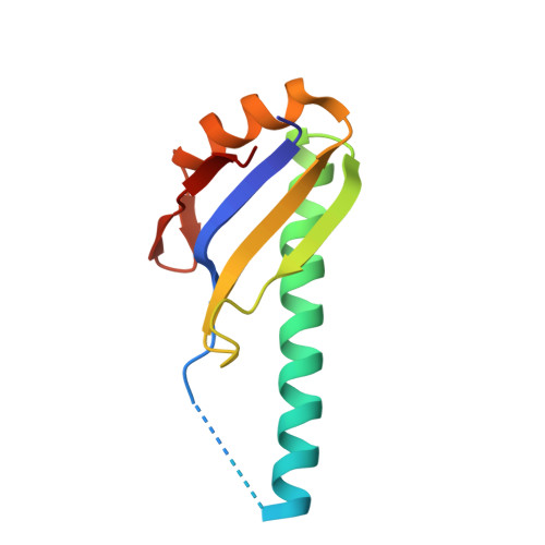
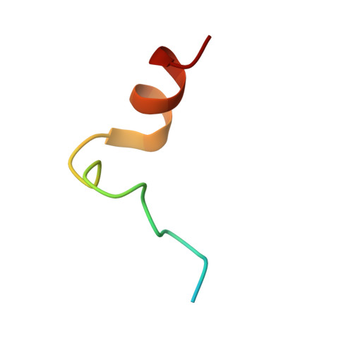
    U2 auxiliary factor (U2AF) is an essential splicing factor that recognizes the 3' splice site and recruits the U2 snRNP to the branch point. The X-ray structure of the human core U2AF heterodimer, consisting of the U2AF35 central domain and a proline-rich region of U2AF65, has been determined at 2.2 A resolution. The structure reveals a novel protein-protein recognition strategy, in which an atypical RNA recognition motif (RRM) of U2AF35 and the U2AF65 polyproline segment interact via reciprocal "tongue-in-groove" tryptophan residues. Complementary biochemical experiments demonstrate that the core U2AF heterodimer binds RNA, and that the interacting tryptophan side chains are essential for U2AF dimerization. Atypical RRMs in other splicing factors may serve as protein-protein interaction motifs elsewhere during spliceosome assembly.


  • Organizational Affiliation

    Laboratories of Molecular Biophysics, The Rockefeller University, 1230 York Avenue, New York, NY 10021, USA.


Macromolecules
Find similar proteins by:  (by identity cutoff)  |  3D Structure
Entity ID: 1
MoleculeChains Sequence LengthOrganismDetailsImage
SPLICING FACTOR U2AF 35 KDA SUBUNIT104Homo sapiensMutation(s): 1 
UniProt & NIH Common Fund Data Resources
Find proteins for Q01081 (Homo sapiens)
Explore Q01081 
Go to UniProtKB:  Q01081
PHAROS:  Q01081
GTEx:  ENSG00000160201 
Entity Groups  
Sequence Clusters30% Identity50% Identity70% Identity90% Identity95% Identity100% Identity
UniProt GroupQ01081
Sequence Annotations
Expand
  • Reference Sequence
Find similar proteins by:  (by identity cutoff)  |  3D Structure
Entity ID: 2
MoleculeChains Sequence LengthOrganismDetailsImage
SPLICING FACTOR U2AF 65 KDA SUBUNIT28Homo sapiensMutation(s): 0 
UniProt & NIH Common Fund Data Resources
Find proteins for P26368 (Homo sapiens)
Explore P26368 
Go to UniProtKB:  P26368
PHAROS:  P26368
GTEx:  ENSG00000063244 
Entity Groups  
Sequence Clusters30% Identity50% Identity70% Identity90% Identity95% Identity100% Identity
UniProt GroupP26368
Sequence Annotations
Expand
  • Reference Sequence
Small Molecules
Ligands 1 Unique
IDChains Name / Formula / InChI Key2D Diagram3D Interactions
HEZ
Query on HEZ

Download Ideal Coordinates CCD File 
C [auth A],
D [auth B]
HEXANE-1,6-DIOL
C6 H14 O2
XXMIOPMDWAUFGU-UHFFFAOYSA-N
Experimental Data & Validation

Experimental Data

  • Method: X-RAY DIFFRACTION
  • Resolution: 2.20 Å
  • R-Value Free: 0.238 
  • R-Value Work: 0.226 
  • R-Value Observed: 0.226 
  • Space Group: P 2 2 2
Unit Cell:
Length ( Å )Angle ( ˚ )
a = 41.52α = 90
b = 49.74β = 90
c = 92.16γ = 90
Software Package:
Software NamePurpose
SOLVEphasing
CNSrefinement
DENZOdata reduction
SCALEPACKdata scaling

Structure Validation

View Full Validation Report


Entry History 

Deposition Data

Revision History  (Full details and data files)

  • Version 1.0: 2001-09-19
    Type: Initial release
  • Version 1.1: 2008-04-27
    Changes: Version format compliance
  • Version 1.2: 2011-07-13
    Changes: Version format compliance
  • Version 1.3: 2021-02-03
    Changes: Database references, Derived calculations, Structure summary
  • Version 1.4: 2021-10-27
    Changes: Database references
  • Version 1.5: 2024-02-07
    Changes: Data collection