


Crystal Structure Analysis of the Dok-5 PTB Domain
Shi, N., Ding, Y., Qiang, B.Q., Yuan, J.G., Rao, Z.H.To be published.
Experimental Data Snapshot
wwPDB Validation 3D Report Full Report

Entity ID: 1 | |||||
|---|---|---|---|---|---|
| Molecule | Chains | Sequence Length | Organism | Details | Image |
| downstream of tyrosine kinase 5 | 103 | Homo sapiens | Mutation(s): 2 Gene Names: DOK5 EC: 2.7.1.112 | 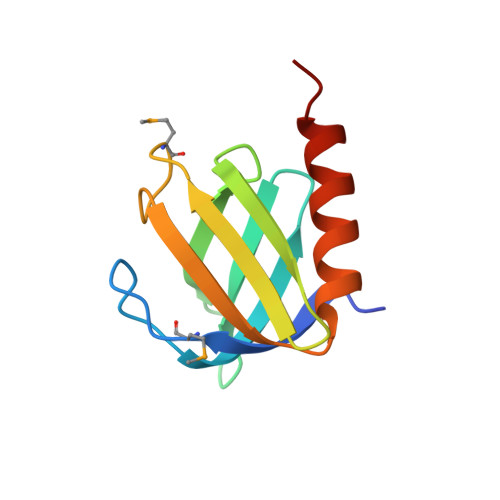 | |
UniProt & NIH Common Fund Data Resources | |||||
Find proteins for Q9P104 (Homo sapiens) Explore Q9P104 Go to UniProtKB: Q9P104 | |||||
PHAROS: Q9P104 GTEx: ENSG00000101134 | |||||
Entity Groups | |||||
| Sequence Clusters | 30% Identity50% Identity70% Identity90% Identity95% Identity100% Identity | ||||
| UniProt Group | Q9P104 | ||||
Sequence AnnotationsExpand | |||||
| |||||
| Modified Residues 1 Unique | |||||
|---|---|---|---|---|---|
| ID | Chains | Type | Formula | 2D Diagram | Parent |
| MSE Query on MSE | A, B | L-PEPTIDE LINKING | C5 H11 N O2 Se |  | MET |
| Length ( Å ) | Angle ( ˚ ) |
|---|---|
| a = 75.89 | α = 90 |
| b = 75.89 | β = 90 |
| c = 107.96 | γ = 120 |
| Software Name | Purpose |
|---|---|
| DENZO | data reduction |
| SCALEPACK | data scaling |
| CNS | refinement |
| CNS | phasing |
RCSB PDB (citation) is hosted by
RCSB PDB is a member of the

