1G50

CRYSTAL STRUCTURE OF A WILD TYPE HER ALPHA LBD AT 2.9 ANGSTROM RESOLUTION


Experimental Data Snapshot

  • Method: X-RAY DIFFRACTION
  • Resolution: 2.90 Å
  • R-Value Free: 0.310 
  • R-Value Work: 0.241 
  • R-Value Observed: 0.241 

wwPDB Validation   3D Report Full Report


Ligand Structure Quality Assessment 


This is version 1.3 of the entry. See complete history


Literature

Overexpression, Purification, and Crystal Structure of Native ER alpha LBD

Eiler, S.Gangloff, M.Duclaud, S.Moras, D.Ruff, M.

(2001) Protein Expr Purif 22: 165-173

  • DOI: https://doi.org/10.1006/prep.2001.1409
  • Primary Citation of Related Structures:  
    1G50

  • PubMed Abstract: 

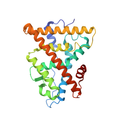
    Several crystal structures of human estrogen receptor alpha ligand-binding domain (hERalpha LBD) complexed with agonist or antagonist molecules have previously been solved. The proteins had been modified in cysteine residues (carboxymethylation) or renatured in urea to circumvent aggregation and denaturation problems. In this work, high-level protein expression and purification together with crystallization screening procedure yielded high amounts of soluble protein without renaturation or modifications steps. The native protein crystallizes in the space group P3(2) 21 with three molecules in the asymmetric unit. The overall structure is very similar to that previously reported for the hERalpha LBD with cysteine carboxymethylated residues thus validating the modification approach. The present strategy can be adapted to other cases where the solubility and the proper folding is a difficulty.


  • Organizational Affiliation

    Laboratoire de Biologie et Génomique Structurales 1, IGBMC, rue Laurent Fries, Illkirch, 67404, France.


Macromolecules
Find similar proteins by:  (by identity cutoff)  |  3D Structure
Entity ID: 1
MoleculeChains Sequence LengthOrganismDetailsImage
ESTROGEN RECEPTOR
A, B, C
247Homo sapiensMutation(s): 0 
UniProt & NIH Common Fund Data Resources
Find proteins for P03372 (Homo sapiens)
Explore P03372 
Go to UniProtKB:  P03372
PHAROS:  P03372
GTEx:  ENSG00000091831 
Entity Groups  
Sequence Clusters30% Identity50% Identity70% Identity90% Identity95% Identity100% Identity
UniProt GroupP03372
Sequence Annotations
Expand
  • Reference Sequence
Small Molecules
Binding Affinity Annotations 
IDSourceBinding Affinity
EST BindingDB:  1G50 Ki: min: 0.11, max: 100 (nM) from 13 assay(s)
Kd: min: 0.2, max: 100 (nM) from 4 assay(s)
IC50: min: 1.00e-2, max: 46 (nM) from 46 assay(s)
EC50: min: 4.00e-3, max: 10 (nM) from 52 assay(s)
PDBBind:  1G50 Kd: 530 (nM) from 1 assay(s)
Experimental Data & Validation

Experimental Data

  • Method: X-RAY DIFFRACTION
  • Resolution: 2.90 Å
  • R-Value Free: 0.310 
  • R-Value Work: 0.241 
  • R-Value Observed: 0.241 
  • Space Group: P 32 2 1
Unit Cell:
Length ( Å )Angle ( ˚ )
a = 105.5α = 90
b = 105.5β = 90
c = 136.08γ = 120
Software Package:
Software NamePurpose
AMoREphasing
CNSrefinement
DENZOdata reduction
SCALEPACKdata scaling

Structure Validation

View Full Validation Report


Ligand Structure Quality Assessment 


Entry History 

Deposition Data

Revision History  (Full details and data files)

  • Version 1.0: 2002-02-06
    Type: Initial release
  • Version 1.1: 2008-04-27
    Changes: Version format compliance
  • Version 1.2: 2011-07-13
    Changes: Version format compliance
  • Version 1.3: 2023-08-09
    Changes: Data collection, Database references, Derived calculations, Refinement description