1FIU

TETRAMERIC RESTRICTION ENDONUCLEASE NGOMIV IN COMPLEX WITH CLEAVED DNA


Experimental Data Snapshot

  • Method: X-RAY DIFFRACTION
  • Resolution: 1.60 Å
  • R-Value Free: 0.204 
  • R-Value Work: 0.173 

wwPDB Validation   3D Report Full Report


This is version 1.3 of the entry. See complete history


Literature

Structure of the tetrameric restriction endonuclease NgoMIV in complex with cleaved DNA.

Deibert, M.Grazulis, S.Sasnauskas, G.Siksnys, V.Huber, R.

(2000) Nat Struct Biol 7: 792-799

  • DOI: https://doi.org/10.1038/79032
  • Primary Citation of Related Structures:  
    1FIU

  • PubMed Abstract: 

    The crystal structure of the NgoMIV restriction endonuclease in complex with cleaved DNA has been determined at 1.6 A resolution. The crystallographic asymmetric unit contains a protein tetramer and two DNA molecules cleaved at their recognition sites. This is the first structure of a tetrameric restriction enzyme-DNA complex. In the tetramer, two primary dimers are arranged back to back with two oligonucleotides bound in clefts on opposite sides of the tetramer. The DNA molecules retain a B-type conformation and have an enclosed angle between their helical axes of 60 degrees. Sequence-specific interactions occur in both the major and minor grooves. Two Mg2+ ions are located close to the cleaved phosphate at the active site of NgoMIV. Biochemical experiments show that interactions between the recognition sites within the tetramer greatly increase DNA cleavage efficiency.


  • Organizational Affiliation

    Max-Planck-Institut für Biochemie, D-82152 Planegg-Martinsried, Germany. markus.deibert@cii.de


Macromolecules

Find similar proteins by:  (by identity cutoff)  |  3D Structure
Entity ID: 3
MoleculeChains Sequence LengthOrganismDetailsImage
TYPE II RESTRICTION ENZYME NGOMII [auth A],
J [auth B],
K [auth C],
L [auth D]
286Neisseria gonorrhoeaeMutation(s): 0 
EC: 3.1.21.4
UniProt
Find proteins for P31032 (Neisseria gonorrhoeae)
Explore P31032 
Go to UniProtKB:  P31032
Entity Groups  
Sequence Clusters30% Identity50% Identity70% Identity90% Identity95% Identity100% Identity
UniProt GroupP31032
Sequence Annotations
Expand
  • Reference Sequence

Find similar nucleic acids by:  Sequence   |   3D Structure  

Entity ID: 1
MoleculeChains LengthOrganismImage
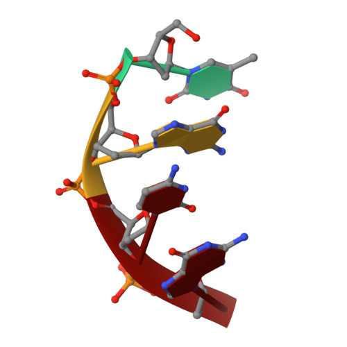
DNA (5'-D(*TP*GP*CP*G)-3')A [auth E],
C [auth F],
E [auth G],
G [auth H]
4N/A
Sequence Annotations
Expand
  • Reference Sequence

Find similar nucleic acids by:  Sequence   |   3D Structure  

Entity ID: 2
MoleculeChains LengthOrganismImage
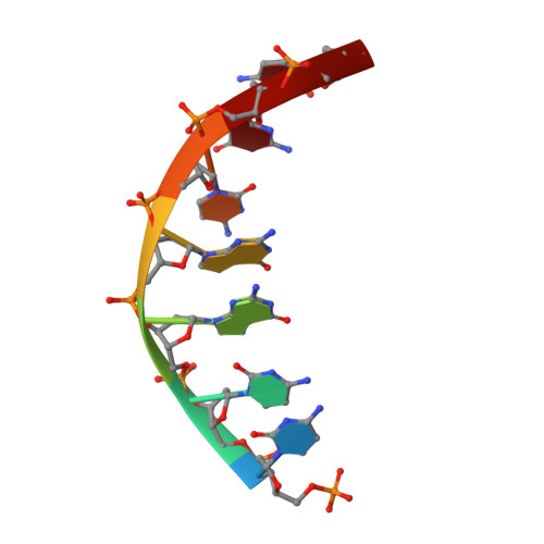
DNA (5'-D(P*CP*CP*GP*GP*CP*GP*C)-3')B [auth I],
D [auth J],
F [auth K],
H [auth L]
7N/A
Sequence Annotations
Expand
  • Reference Sequence
Small Molecules
Experimental Data & Validation

Experimental Data

  • Method: X-RAY DIFFRACTION
  • Resolution: 1.60 Å
  • R-Value Free: 0.204 
  • R-Value Work: 0.173 
  • Space Group: P 21 21 21
Unit Cell:
Length ( Å )Angle ( ˚ )
a = 90.4α = 90
b = 91.13β = 90
c = 149.52γ = 90
Software Package:
Software NamePurpose
SHARPphasing
X-PLORrefinement
DENZOdata reduction
SCALEPACKdata scaling

Structure Validation

View Full Validation Report


Entry History 

Deposition Data

Revision History  (Full details and data files)

  • Version 1.0: 2001-02-07
    Type: Initial release
  • Version 1.1: 2008-04-27
    Changes: Version format compliance
  • Version 1.2: 2011-07-13
    Changes: Version format compliance
  • Version 1.3: 2024-02-07
    Changes: Data collection, Database references, Derived calculations