1F1J

CRYSTAL STRUCTURE OF CASPASE-7 IN COMPLEX WITH ACETYL-ASP-GLU-VAL-ASP-CHO


Experimental Data Snapshot

  • Method: X-RAY DIFFRACTION
  • Resolution: 2.35 Å
  • R-Value Free: 0.263 
  • R-Value Work: 0.185 
  • R-Value Observed: 0.185 

wwPDB Validation   3D Report Full Report


This is version 1.4 of the entry. See complete history


Literature

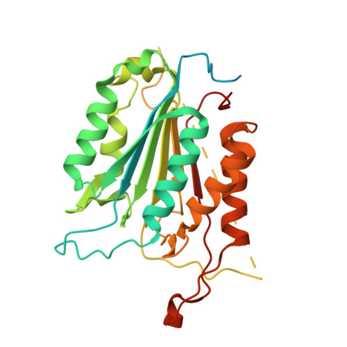
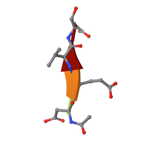
The structures of caspases-1, -3, -7 and -8 reveal the basis for substrate and inhibitor selectivity.

Wei, Y.Fox, T.Chambers, S.P.Sintchak, J.Coll, J.T.Golec, J.M.Swenson, L.Wilson, K.P.Charifson, P.S.

(2000) Chem Biol 7: 423-432

  • DOI: https://doi.org/10.1016/s1074-5521(00)00123-x
  • Primary Citation of Related Structures:  
    1F1J

  • PubMed Abstract: 

    Peptide inhibitors of caspases have helped define the role of these cysteine proteases in biology. Structural and biochemical characterization of the caspase enzymes may contribute to the development of new drugs for the treatment of caspase-mediated inflammation and apoptosis.


  • Organizational Affiliation

    Vertex Pharmaceuticals, Cambridge, MA 02139-4242, USA.


Macromolecules
Find similar proteins by:  (by identity cutoff)  |  3D Structure
Entity ID: 1
MoleculeChains Sequence LengthOrganismDetailsImage
CASPASE-7 PROTEASE
A, B
305Homo sapiensMutation(s): 0 
EC: 3.4.22
UniProt & NIH Common Fund Data Resources
Find proteins for P55210 (Homo sapiens)
Explore P55210 
Go to UniProtKB:  P55210
PHAROS:  P55210
GTEx:  ENSG00000165806 
Entity Groups  
Sequence Clusters30% Identity50% Identity70% Identity90% Identity95% Identity100% Identity
UniProt GroupP55210
Sequence Annotations
Expand
  • Reference Sequence

Find similar proteins by:  Sequence   |   3D Structure  

Entity ID: 2
MoleculeChains Sequence LengthOrganismDetailsImage
ACE-ASP-GLU-VAL-ASP-CHO
C, D
5N/AMutation(s): 0 
Sequence Annotations
Expand
  • Reference Sequence
Small Molecules
Ligands 1 Unique
IDChains Name / Formula / InChI Key2D Diagram3D Interactions
SO4
Query on SO4

Download Ideal Coordinates CCD File 
E [auth A],
F [auth B]
SULFATE ION
O4 S
QAOWNCQODCNURD-UHFFFAOYSA-L
Biologically Interesting Molecules (External Reference) 1 Unique
Experimental Data & Validation

Experimental Data

  • Method: X-RAY DIFFRACTION
  • Resolution: 2.35 Å
  • R-Value Free: 0.263 
  • R-Value Work: 0.185 
  • R-Value Observed: 0.185 
  • Space Group: P 32 2 1
Unit Cell:
Length ( Å )Angle ( ˚ )
a = 88.18α = 90
b = 88.18β = 90
c = 186.23γ = 120
Software Package:
Software NamePurpose
AMoREphasing
X-PLORrefinement
DENZOdata reduction
SCALEPACKdata scaling

Structure Validation

View Full Validation Report


Entry History 

Deposition Data

Revision History  (Full details and data files)

  • Version 1.0: 2001-05-23
    Type: Initial release
  • Version 1.1: 2008-04-27
    Changes: Version format compliance
  • Version 1.2: 2011-07-13
    Changes: Atomic model, Database references, Derived calculations, Non-polymer description, Structure summary, Version format compliance
  • Version 1.3: 2012-12-12
    Changes: Other
  • Version 1.4: 2013-07-03
    Changes: Structure summary