1EMU

STRUCTURE OF THE AXIN RGS-HOMOLOGOUS DOMAIN IN COMPLEX WITH A SAMP REPEAT FROM APC


Experimental Data Snapshot

  • Method: X-RAY DIFFRACTION
  • Resolution: 1.90 Å
  • R-Value Free: 0.221 
  • R-Value Work: 0.205 
  • R-Value Observed: 0.205 

wwPDB Validation   3D Report Full Report


This is version 1.3 of the entry. See complete history


Literature

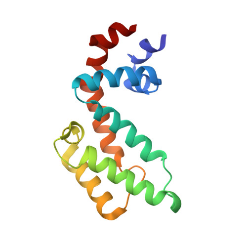
Structural basis of the Axin-adenomatous polyposis coli interaction.

Spink, K.E.Polakis, P.Weis, W.I.

(2000) EMBO J 19: 2270-2279

  • DOI: https://doi.org/10.1093/emboj/19.10.2270
  • Primary Citation of Related Structures:  
    1DK8, 1EMU

  • PubMed Abstract: 

    Axin and the adenomatous polyposis coli (APC) tumor suppressor protein are components of the Wnt/Wingless growth factor signaling pathway. In the absence of Wnt signal, Axin and APC regulate cytoplasmic levels of the proto-oncogene beta-catenin through the formation of a large complex containing these three proteins, glycogen synthase kinase 3beta (GSK3beta) and several other proteins. Both Axin and APC are known to be critical for beta-catenin regulation, and truncations in APC that eliminate the Axin-binding site result in human cancers. A protease-resistant domain of Axin that contains the APC-binding site is a member of the regulators of G-protein signaling (RGS) superfamily. The crystal structures of this domain alone and in complex with an Axin-binding sequence from APC reveal that the Axin-APC interaction occurs at a conserved groove on a face of the protein that is distinct from the G-protein interface of classical RGS proteins. The molecular interactions observed in the Axin-APC complex provide a rationale for the evolutionary conservation seen in both proteins.


  • Organizational Affiliation

    Department of Structural Biology, Stanford University School of Medicine, 299 Campus Drive West, Stanford, CA 94305, USA.


Macromolecules
Find similar proteins by:  (by identity cutoff)  |  3D Structure
Entity ID: 1
MoleculeChains Sequence LengthOrganismDetailsImage
AXIN132Homo sapiensMutation(s): 0 
UniProt & NIH Common Fund Data Resources
Find proteins for O15169 (Homo sapiens)
Explore O15169 
Go to UniProtKB:  O15169
PHAROS:  O15169
GTEx:  ENSG00000103126 
Entity Groups  
Sequence Clusters30% Identity50% Identity70% Identity90% Identity95% Identity100% Identity
UniProt GroupO15169
Sequence Annotations
Expand
  • Reference Sequence

Find similar proteins by:  Sequence   |   3D Structure  

Entity ID: 2
MoleculeChains Sequence LengthOrganismDetailsImage
ADENOMATOUS POLYPOSIS COLI PROTEIN16N/AMutation(s): 0 
UniProt & NIH Common Fund Data Resources
Find proteins for P25054 (Homo sapiens)
Explore P25054 
Go to UniProtKB:  P25054
PHAROS:  P25054
GTEx:  ENSG00000134982 
Entity Groups  
Sequence Clusters30% Identity50% Identity70% Identity90% Identity95% Identity100% Identity
UniProt GroupP25054
Sequence Annotations
Expand
  • Reference Sequence
Experimental Data & Validation

Experimental Data

  • Method: X-RAY DIFFRACTION
  • Resolution: 1.90 Å
  • R-Value Free: 0.221 
  • R-Value Work: 0.205 
  • R-Value Observed: 0.205 
  • Space Group: P 21 21 21
Unit Cell:
Length ( Å )Angle ( ˚ )
a = 30.6α = 90
b = 69.98β = 90
c = 71.96γ = 90
Software Package:
Software NamePurpose
CNSrefinement
DENZOdata reduction
SCALEPACKdata scaling
CNSphasing

Structure Validation

View Full Validation Report


Entry History 

Deposition Data

Revision History  (Full details and data files)

  • Version 1.0: 2000-07-05
    Type: Initial release
  • Version 1.1: 2008-04-27
    Changes: Version format compliance
  • Version 1.2: 2011-07-13
    Changes: Non-polymer description, Version format compliance
  • Version 1.3: 2024-02-07
    Changes: Data collection, Database references, Derived calculations