1CWE

HUMAN P56LCK TYROSINE KINASE COMPLEXED WITH PHOSPHOPEPTIDE


Experimental Data Snapshot

  • Method: X-RAY DIFFRACTION
  • Resolution: 2.30 Å
  • R-Value Work: 0.192 
  • R-Value Observed: 0.192 

wwPDB Validation   3D Report Full Report


This is version 1.4 of the entry. See complete history


Literature

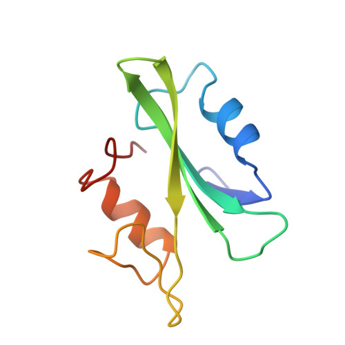
The crystal structures of the SH2 domain of p56lck complexed with two phosphopeptides suggest a gated peptide binding site.

Mikol, V.Baumann, G.Keller, T.H.Manning, U.Zurini, M.G.

(1995) J Mol Biol 246: 344-355

  • DOI: https://doi.org/10.1006/jmbi.1994.0089
  • Primary Citation of Related Structures:  
    1CWD, 1CWE

  • PubMed Abstract: 

    Src homology-2 (SH2) domains are protein modules found within a wide variety of cytoplasmic signalling molecules that bind with high affinity to phosphotyrosyl-containing protein sequences. In order to develop SH2 inhibitors that contain phosphotyrosyl analogues resistant to cellular phosphatases, we have solved the crystal structures of the SH2 domain of p56lck in separate complexes with two high-affinity p-(phosphonomethyl)phenylalanine-containing peptides. The structures have been determined at 2.3 A and 2.25 A, and refined to crystallographic R-factors of 19.2% and 18.5%, respectively. The conformation of the SH2 domain of p56lck is essentially similar to that observed in Src and Lck complexed with a phosphotyrosine-containing peptide except in some loops and especially in the loop that connects the second and third beta-strands. This loop, which was involved in hydrogen-bond interactions with the phosphotyrosine moiety, has moved away in the phosphonopeptide complexes as a rigid body by about 7 A on two hinges leaving the tyrosine phosphate mimetic moiety accessible to the solvent. Some intramolecular hydrogen bonds with other residues of the third and fourth beta-strands stabilize an open conformation of the lid, suggesting a flap mechanism for peptide binding.


  • Organizational Affiliation

    Preclinical Research, Sandoz Pharma AG, CH-4002, Basel, Switzerland.


Macromolecules
Find similar proteins by:  (by identity cutoff)  |  3D Structure
Entity ID: 1
MoleculeChains Sequence LengthOrganismDetailsImage
P56LCK TYROSINE KINASE
A, C
98Homo sapiensMutation(s): 0 
EC: 2.7.1.112
UniProt & NIH Common Fund Data Resources
Find proteins for P06239 (Homo sapiens)
Explore P06239 
Go to UniProtKB:  P06239
PHAROS:  P06239
GTEx:  ENSG00000182866 
Entity Groups  
Sequence Clusters30% Identity50% Identity70% Identity90% Identity95% Identity100% Identity
UniProt GroupP06239
Sequence Annotations
Expand
  • Reference Sequence

Find similar proteins by:  Sequence   |   3D Structure  

Entity ID: 2
MoleculeChains Sequence LengthOrganismDetailsImage
PHOSPHOPEPTIDE ACQ-PMP-GLU-GLU-ILE-PRO
B, D
7synthetic constructMutation(s): 0 
Sequence Annotations
Expand
  • Reference Sequence
Small Molecules
Modified Residues  1 Unique
IDChains TypeFormula2D DiagramParent
PM3
Query on PM3
B, D
L-PEPTIDE LINKINGC10 H14 N O5 PPHE
Experimental Data & Validation

Experimental Data

  • Method: X-RAY DIFFRACTION
  • Resolution: 2.30 Å
  • R-Value Work: 0.192 
  • R-Value Observed: 0.192 
  • Space Group: P 1
Unit Cell:
Length ( Å )Angle ( ˚ )
a = 35.17α = 68.71
b = 41.84β = 82.94
c = 45.52γ = 87.36
Software Package:
Software NamePurpose
X-PLORrefinement

Structure Validation

View Full Validation Report


Entry History 

Deposition Data

  • Released Date: 1996-12-07 
  • Deposition Author(s): Mikol, V.

Revision History  (Full details and data files)

  • Version 1.0: 1996-12-07
    Type: Initial release
  • Version 1.1: 2008-03-24
    Changes: Version format compliance
  • Version 1.2: 2011-07-13
    Changes: Version format compliance
  • Version 1.3: 2011-08-03
    Changes: Structure summary
  • Version 1.4: 2011-08-17
    Changes: Source and taxonomy