1AN2

RECOGNITION BY MAX OF ITS COGNATE DNA THROUGH A DIMERIC B/HLH/Z DOMAIN


Experimental Data Snapshot

  • Method: X-RAY DIFFRACTION
  • Resolution: 2.90 Å
  • R-Value Work: 0.232 
  • R-Value Observed: 0.232 

wwPDB Validation   3D Report Full Report


This is version 1.5 of the entry. See complete history


Literature

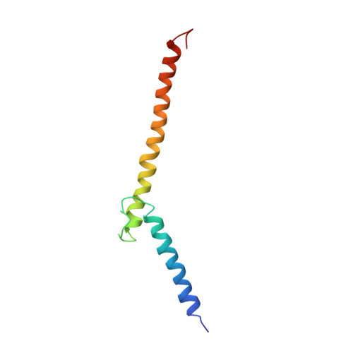
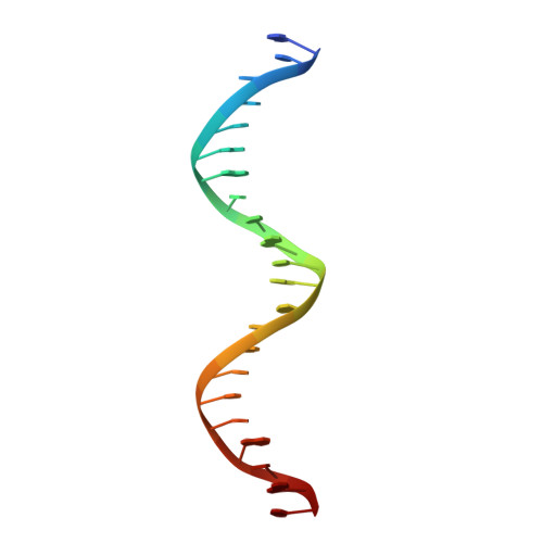
Recognition by Max of its cognate DNA through a dimeric b/HLH/Z domain.

Ferre-D'Amare, A.R.Prendergast, G.C.Ziff, E.B.Burley, S.K.

(1993) Nature 363: 38-45

  • DOI: https://doi.org/10.1038/363038a0
  • Primary Citation of Related Structures:  
    1AN2

  • PubMed Abstract: 

    The three-dimensional structure of the basic/helix-loop-helix/leucine zipper domain of the transcription factor Max complexed with DNA has been determined by X-ray crystallography at 2.9 A resolution. Max binds as a dimer to its recognition sequence CACGTG by direct contacts between the alpha-helical basic region and the major groove. This symmetric homodimer, a new protein fold, is a parallel, left-handed, four-helix bundle, with each monomer containing two alpha-helical segments separated by a loop. The two alpha-helical segments are composed of the basic region plus helix 1 and helix 2 plus the leucine repeat, respectively. As in GCN4, the leucine repeat forms a parallel coiled coil.


  • Organizational Affiliation

    Laboratories of Molecular Biophysics, Rockefeller University, New York, New York 10021.


Macromolecules

Find similar proteins by:  (by identity cutoff)  |  3D Structure
Entity ID: 2
MoleculeChains Sequence LengthOrganismDetailsImage
PROTEIN (TRANSCRIPTION FACTOR MAX (TF MAX))B [auth A]86Homo sapiensMutation(s): 0 
Gene Names: MAX
UniProt & NIH Common Fund Data Resources
Find proteins for P61244 (Homo sapiens)
Explore P61244 
Go to UniProtKB:  P61244
PHAROS:  P61244
GTEx:  ENSG00000125952 
Entity Groups  
Sequence Clusters30% Identity50% Identity70% Identity90% Identity95% Identity100% Identity
UniProt GroupP61244
Sequence Annotations
Expand
  • Reference Sequence

Find similar nucleic acids by:  Sequence   |   3D Structure  

Entity ID: 1
MoleculeChains LengthOrganismImage
DNA (5'-D(*GP*TP*GP*TP*AP*GP*GP*TP*CP*AP*CP*GP*TP*GP*AP*CP*C P*TP*AP*CP*AP*C)- 3')A [auth B]22Mus musculus
Sequence Annotations
Expand
  • Reference Sequence
Experimental Data & Validation

Experimental Data

  • Method: X-RAY DIFFRACTION
  • Resolution: 2.90 Å
  • R-Value Work: 0.232 
  • R-Value Observed: 0.232 
  • Space Group: P 61 2 2
Unit Cell:
Length ( Å )Angle ( ˚ )
a = 72.2α = 90
b = 72.2β = 90
c = 146.4γ = 120
Software Package:
Software NamePurpose
X-PLORrefinement
DENZOdata reduction
SCALEPACKdata scaling

Structure Validation

View Full Validation Report


Entry History 

Deposition Data

Revision History  (Full details and data files)

  • Version 1.0: 1997-09-17
    Type: Initial release
  • Version 1.1: 2008-05-22
    Changes: Version format compliance
  • Version 1.2: 2011-07-13
    Changes: Version format compliance
  • Version 1.3: 2013-04-03
    Changes: Version format compliance
  • Version 1.4: 2021-02-03
    Changes: Database references, Structure summary
  • Version 1.5: 2024-02-07
    Changes: Data collection, Database references