1AIN

CRYSTAL STRUCTURE OF HUMAN ANNEXIN I AT 2.5 ANGSTROMS RESOLUTION


Experimental Data Snapshot

  • Method: X-RAY DIFFRACTION
  • Resolution: 2.50 Å
  • R-Value Work: 0.163 

wwPDB Validation   3D Report Full Report


This is version 1.3 of the entry. See complete history


Literature

Crystal structure of human annexin I at 2.5 A resolution.

Weng, X.Luecke, H.Song, I.S.Kang, D.S.Kim, S.H.Huber, R.

(1993) Protein Sci 2: 448-458

  • DOI: https://doi.org/10.1002/pro.5560020317
  • Primary Citation of Related Structures:  
    1AIN

  • PubMed Abstract: 

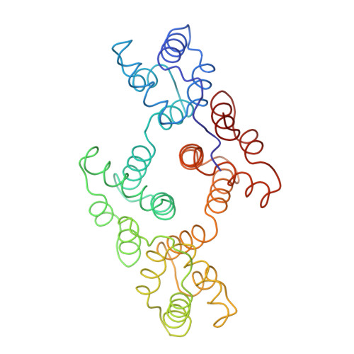
    cDNA coding for N-terminally truncated human annexin I, a member of the family of Ca(2+)-dependent phospholipid binding proteins, has been cloned and expressed in Escherichia coli. The expressed protein is biologically active, and has been purified and crystallized in space group P2(1)2(1)2(1) with cell dimensions a = 139.36 A, b = 67.50 A, and c = 42.11 A. The crystal structure has been determined by molecular replacement at 3.0 A resolution using the annexin V core structure as the search model. The average backbone deviation between these two structures is 2.34 A. The structure has been refined to an R-factor of 17.7% at 2.5 A resolution. Six calcium sites have been identified in the annexin I structure. Each is located in the loop region of the helix-loop-helix motif. Two of the six calcium sites in annexin I are not occupied in the annexin V structure. The superpositions of the corresponding loop regions in the four domains show that the calcium binding loops in annexin I can be divided into two classes: type II and type III. Both classes are different from the well-known EF-hand motif (type I).


  • Organizational Affiliation

    Department of Molecular and Cell Biology, University of California, Berkeley 94720.


Macromolecules
Find similar proteins by:  (by identity cutoff)  |  3D Structure
Entity ID: 1
MoleculeChains Sequence LengthOrganismDetailsImage
ANNEXIN I314Homo sapiensMutation(s): 0 
UniProt & NIH Common Fund Data Resources
Find proteins for P04083 (Homo sapiens)
Explore P04083 
Go to UniProtKB:  P04083
PHAROS:  P04083
GTEx:  ENSG00000135046 
Entity Groups  
Sequence Clusters30% Identity50% Identity70% Identity90% Identity95% Identity100% Identity
UniProt GroupP04083
Sequence Annotations
Expand
  • Reference Sequence
Experimental Data & Validation

Experimental Data

  • Method: X-RAY DIFFRACTION
  • Resolution: 2.50 Å
  • R-Value Work: 0.163 
  • Space Group: P 21 21 21
Unit Cell:
Length ( Å )Angle ( ˚ )
a = 139.36α = 90
b = 67.5β = 90
c = 42.11γ = 90
Software Package:
Software NamePurpose
X-PLORmodel building
TNTrefinement
X-PLORphasing

Structure Validation

View Full Validation Report


Entry History 

Deposition Data

  • Released Date: 1993-07-15 
  • Deposition Author(s): Kim, S.-H.

Revision History  (Full details and data files)

  • Version 1.0: 1993-07-15
    Type: Initial release
  • Version 1.1: 2008-03-03
    Changes: Version format compliance
  • Version 1.2: 2011-07-13
    Changes: Version format compliance
  • Version 1.3: 2024-02-07
    Changes: Data collection, Database references, Other