8TYQ

Structure of the C-terminal half of LRRK2 bound to GZD-824 (G2019S mutant)


Experimental Data Snapshot

  • Method: ELECTRON MICROSCOPY
  • Resolution: 2.99 Å
  • Aggregation State: PARTICLE 
  • Reconstruction Method: SINGLE PARTICLE 

wwPDB Validation   3D Report Full Report


This is version 1.1 of the entry. See complete history


Literature

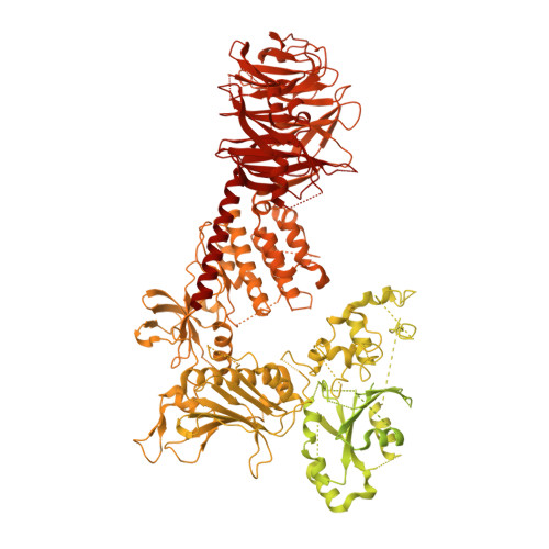
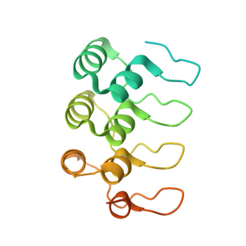
Inhibition of Parkinson's disease-related LRRK2 by type I and type II kinase inhibitors: Activity and structures.

Sanz Murillo, M.Villagran Suarez, A.Dederer, V.Chatterjee, D.Alegrio Louro, J.Knapp, S.Mathea, S.Leschziner, A.E.

(2023) Sci Adv 9: eadk6191-eadk6191

  • DOI: https://doi.org/10.1126/sciadv.adk6191
  • Primary Citation of Related Structures:  
    8TXZ, 8TYQ, 8TZB, 8TZC, 8TZE, 8TZF, 8TZG, 8TZH

  • PubMed Abstract: 

    Mutations in leucine-rich repeat kinase 2 (LRRK2) are a common cause of familial Parkinson's disease (PD) and a risk factor for the sporadic form. Increased kinase activity was shown in patients with both familial and sporadic PD, making LRRK2 kinase inhibitors a major focus of drug development efforts. Although much progress has been made in understanding the structural biology of LRRK2, there are no available structures of LRRK2 inhibitor complexes. To this end, we solved cryo-electron microscopy structures of LRRK2, wild-type and PD-linked mutants, bound to the LRRK2-specific type I inhibitor MLi-2 and the broad-spectrum type II inhibitor GZD-824. Our structures revealed an active-like LRRK2 kinase in the type I inhibitor complex, and an inactive DYG-out in the type II inhibitor complex. Our structural analysis also showed how inhibitor-induced conformational changes in LRRK2 are affected by its autoinhibitory N-terminal repeats. The structures provide a template for the rational development of LRRK2 kinase inhibitors covering both canonical inhibitor binding modes.


  • Organizational Affiliation

    Department of Cellular and Molecular Medicine, School of Medicine, University of California San Diego, La Jolla, CA 92093, USA.


Macromolecules
Find similar proteins by:  (by identity cutoff)  |  3D Structure
Entity ID: 1
MoleculeChains Sequence LengthOrganismDetailsImage
Leucine-rich repeat serine/threonine-protein kinase 22,527Homo sapiensMutation(s): 1 
Gene Names: LRRK2
UniProt & NIH Common Fund Data Resources
Find proteins for Q5S007 (Homo sapiens)
Explore Q5S007 
Go to UniProtKB:  Q5S007
PHAROS:  Q5S007
GTEx:  ENSG00000188906 
Entity Groups  
Sequence Clusters30% Identity50% Identity70% Identity90% Identity95% Identity100% Identity
UniProt GroupQ5S007
Sequence Annotations
Expand
  • Reference Sequence
Find similar proteins by:  (by identity cutoff)  |  3D Structure
Entity ID: 2
MoleculeChains Sequence LengthOrganismDetailsImage
Designed Ankyrin Repeats Protein E11182synthetic constructMutation(s): 0 
Entity Groups  
Sequence Clusters30% Identity50% Identity70% Identity90% Identity95% Identity100% Identity
Sequence Annotations
Expand
  • Reference Sequence
Small Molecules
Ligands 1 Unique
IDChains Name / Formula / InChI Key2D Diagram3D Interactions
T3X (Subject of Investigation/LOI)
Query on T3X

Download Ideal Coordinates CCD File 
C [auth A]4-methyl-N-{4-[(4-methylpiperazin-1-yl)methyl]-3-(trifluoromethyl)phenyl}-3-[(1H-pyrazolo[3,4-b]pyridin-5-yl)ethynyl]benzamide
C29 H27 F3 N6 O
TZKBVRDEOITLRB-UHFFFAOYSA-N
Experimental Data & Validation

Experimental Data

  • Method: ELECTRON MICROSCOPY
  • Resolution: 2.99 Å
  • Aggregation State: PARTICLE 
  • Reconstruction Method: SINGLE PARTICLE 
EM Software:
TaskSoftware PackageVersion
RECONSTRUCTIONcryoSPARC
MODEL REFINEMENTPHENIX

Structure Validation

View Full Validation Report


Entry History & Funding Information

Deposition Data


Funding OrganizationLocationGrant Number
Michael J. Fox FoundationUnited StatesASAP-000519

Revision History  (Full details and data files)

  • Version 1.0: 2023-12-06
    Type: Initial release
  • Version 1.1: 2023-12-27
    Changes: Database references