8TQ0

Crystal structure of Danio rerio histone deacetylase 6 catalytic domain 2 complexed with (R)-Lipoic Acid


Experimental Data Snapshot

  • Method: X-RAY DIFFRACTION
  • Resolution: 2.40 Å
  • R-Value Free: 0.248 
  • R-Value Work: 0.207 
  • R-Value Observed: 0.210 

wwPDB Validation   3D Report Full Report


Ligand Structure Quality Assessment 


This is version 1.2 of the entry. See complete history


Literature

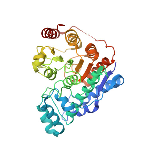
Crystal structure of histone deacetylase 6 complexed with (R)-lipoic acid, an essential cofactor in central carbon metabolism.

Watson, P.R.Stollmaier, J.G.Christianson, D.W.

(2023) J Biol Chem 299: 105228-105228

  • DOI: https://doi.org/10.1016/j.jbc.2023.105228
  • Primary Citation of Related Structures:  
    8TQ0

  • PubMed Abstract: 

    The enzyme cofactor (R)-lipoic acid plays a critical role in central carbon metabolism due to its catalytic function in the generation of acetyl-CoA, which links glycolysis with the tricarboxylic acid cycle. This cofactor is also essential for the generation of succinyl CoA within the tricarboxylic acid cycle. However, the biological functions of (R)-lipoic acid extend beyond metabolism owing to its facile redox chemistry. Most recently, the reduced form of (R)-lipoic acid, (R)-dihydrolipoic acid, has been shown to inhibit histone deacetylases (HDACs) with selectivity for the inhibition of HDAC6. Here, we report the 2.4 Å-resolution X-ray crystal structure of the complex between (R)-dihydrolipoic acid and HDAC6 catalytic domain 2 from Danio rerio, and we report a dissociation constant (K D ) of 350 nM for this complex as determined by isothermal titration calorimetry. The crystal structure illuminates key affinity determinants in the enzyme active site, including thiolate-Zn 2+ coordination and S-π interactions in the F583-F643 aromatic crevice. This study provides the first visualization of the connection between HDAC function and the biological response to oxidative stress: the dithiol moiety of (R)-dihydrolipoic acid can serve as a redox-regulated pharmacophore capable of simultaneously targeting the catalytic Zn 2+ ion and the aromatic crevice in the active site of HDAC6.


  • Organizational Affiliation

    Roy and Diana Vagelos Laboratories, Department of Chemistry, University of Pennsylvania, Philadelphia, Pennsylvania, United States.


Macromolecules
Find similar proteins by:  (by identity cutoff)  |  3D Structure
Entity ID: 1
MoleculeChains Sequence LengthOrganismDetailsImage
Hdac6 protein
A, B
364Danio rerioMutation(s): 0 
Gene Names: hdac6
UniProt
Find proteins for F8W4B7 (Danio rerio)
Explore F8W4B7 
Go to UniProtKB:  F8W4B7
Entity Groups  
Sequence Clusters30% Identity50% Identity70% Identity90% Identity95% Identity100% Identity
UniProt GroupF8W4B7
Sequence Annotations
Expand
  • Reference Sequence
Small Molecules
Ligands 3 Unique
IDChains Name / Formula / InChI Key2D Diagram3D Interactions
RED (Subject of Investigation/LOI)
Query on RED

Download Ideal Coordinates CCD File 
C [auth A],
G [auth B]
DIHYDROLIPOIC ACID
C8 H16 O2 S2
IZFHEQBZOYJLPK-SSDOTTSWSA-N
ZN
Query on ZN

Download Ideal Coordinates CCD File 
D [auth A],
H [auth B]
ZINC ION
Zn
PTFCDOFLOPIGGS-UHFFFAOYSA-N
K
Query on K

Download Ideal Coordinates CCD File 
E [auth A],
F [auth A],
I [auth B],
J [auth B]
POTASSIUM ION
K
NPYPAHLBTDXSSS-UHFFFAOYSA-N
Experimental Data & Validation

Experimental Data

  • Method: X-RAY DIFFRACTION
  • Resolution: 2.40 Å
  • R-Value Free: 0.248 
  • R-Value Work: 0.207 
  • R-Value Observed: 0.210 
  • Space Group: P 21 21 21
Unit Cell:
Length ( Å )Angle ( ˚ )
a = 74.894α = 90
b = 92.156β = 90
c = 96.666γ = 90
Software Package:
Software NamePurpose
PHENIXrefinement
XDSdata reduction
Aimlessdata scaling
PHASERphasing

Structure Validation

View Full Validation Report


Ligand Structure Quality Assessment 


Entry History & Funding Information

Deposition Data


Funding OrganizationLocationGrant Number
National Institutes of Health/National Institute of General Medical Sciences (NIH/NIGMS)United StatesGM49758

Revision History  (Full details and data files)

  • Version 1.0: 2023-09-20
    Type: Initial release
  • Version 1.1: 2023-10-04
    Changes: Structure summary
  • Version 1.2: 2024-04-03
    Changes: Database references