8IF3

Structure of human alpha-2/delta-1 with mirogabalin


Experimental Data Snapshot

  • Method: ELECTRON MICROSCOPY
  • Resolution: 3.23 Å
  • Aggregation State: PARTICLE 
  • Reconstruction Method: SINGLE PARTICLE 

wwPDB Validation   3D Report Full Report


This is version 1.1 of the entry. See complete history


Literature

Recognition Mechanism of a Novel Gabapentinoid Drug, Mirogabalin, for Recombinant Human alpha 2 delta 1, a Voltage-Gated Calcium Channel Subunit.

Kozai, D.Numoto, N.Nishikawa, K.Kamegawa, A.Kawasaki, S.Hiroaki, Y.Irie, K.Oshima, A.Hanzawa, H.Shimada, K.Kitano, Y.Fujiyoshi, Y.

(2023) J Mol Biol 435: 168049-168049

  • DOI: https://doi.org/10.1016/j.jmb.2023.168049
  • Primary Citation of Related Structures:  
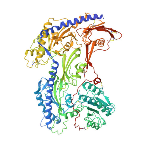
    8IF3, 8IF4

  • PubMed Abstract: 

    Mirogabalin is a novel gabapentinoid drug with a hydrophobic bicyclo substituent on the γ-aminobutyric acid moiety that targets the voltage-gated calcium channel subunit α 2 δ1. Here, to reveal the mirogabalin recognition mechanisms of α 2 δ1, we present structures of recombinant human α 2 δ1 with and without mirogabalin analyzed by cryo-electron microscopy. These structures show the binding of mirogabalin to the previously reported gabapentinoid binding site, which is the extracellular dCache_1 domain containing a conserved amino acid binding motif. A slight conformational change occurs around the residues positioned close to the hydrophobic group of mirogabalin. Mutagenesis binding assays identified that residues in the hydrophobic interaction region, in addition to several amino acid binding motif residues around the amino and carboxyl groups of mirogabalin, are critical for mirogabalin binding. The A215L mutation introduced to decrease the hydrophobic pocket volume predictably suppressed mirogabalin binding and promoted the binding of another ligand, L-Leu, with a smaller hydrophobic substituent than mirogabalin. Alterations of residues in the hydrophobic interaction region of α 2 δ1 to those of the α 2 δ2, α 2 δ3, and α 2 δ4 isoforms, of which α 2 δ3 and α 2 δ4 are gabapentin-insensitive, suppressed the binding of mirogabalin. These results support the importance of hydrophobic interactions in α 2 δ1 ligand recognition.


  • Organizational Affiliation

    Cellular and Structural Physiology Institute (CeSPI), Nagoya University, Furo-cho, Chikusa-ku, Nagoya 464-8601, Japan; Japan Biological Informatics Consortium, 2-4-32 Aomi, Koto-ku, Tokyo 135-0063, Japan; Advanced Research Institute, Tokyo Medical and Dental University, 1-5-45 Yushima, Bunkyo-ku, Tokyo 113-8501, Japan.


Macromolecules
Find similar proteins by:  (by identity cutoff)  |  3D Structure
Entity ID: 1
MoleculeChains Sequence LengthOrganismDetailsImage
Voltage-dependent calcium channel subunit alpha-2/delta-11,099Homo sapiensMutation(s): 0 
Gene Names: CACNA2D1CACNL2ACCHL2AMHS3
Membrane Entity: Yes 
UniProt & NIH Common Fund Data Resources
Find proteins for P54289 (Homo sapiens)
Explore P54289 
Go to UniProtKB:  P54289
PHAROS:  P54289
GTEx:  ENSG00000153956 
Entity Groups  
Sequence Clusters30% Identity50% Identity70% Identity90% Identity95% Identity100% Identity
UniProt GroupP54289
Sequence Annotations
Expand
  • Reference Sequence
Oligosaccharides

Help

Entity ID: 2
MoleculeChains Length2D Diagram Glycosylation3D Interactions
2-acetamido-2-deoxy-beta-D-glucopyranose-(1-4)-2-acetamido-2-deoxy-beta-D-glucopyranoseB [auth C],
C [auth F]
2N-Glycosylation
Glycosylation Resources
GlyTouCan:  G42666HT
GlyCosmos:  G42666HT
GlyGen:  G42666HT
Small Molecules
Ligands 2 Unique
IDChains Name / Formula / InChI Key2D Diagram3D Interactions
NAG
Query on NAG

Download Ideal Coordinates CCD File 
D [auth A]
E [auth A]
F [auth A]
G [auth A]
I [auth A]
D [auth A],
E [auth A],
F [auth A],
G [auth A],
I [auth A],
J [auth A],
K [auth A]
2-acetamido-2-deoxy-beta-D-glucopyranose
C8 H15 N O6
OVRNDRQMDRJTHS-FMDGEEDCSA-N
8X9 (Subject of Investigation/LOI)
Query on 8X9

Download Ideal Coordinates CCD File 
H [auth A]2-[(1R,5S,6S)-6-(aminomethyl)-3-ethyl-6-bicyclo[3.2.0]hept-3-enyl]acetic acid
C12 H19 N O2
FTBQORVNHOIASH-CKYFFXLPSA-N
Experimental Data & Validation

Experimental Data

  • Method: ELECTRON MICROSCOPY
  • Resolution: 3.23 Å
  • Aggregation State: PARTICLE 
  • Reconstruction Method: SINGLE PARTICLE 
EM Software:
TaskSoftware PackageVersion
MODEL REFINEMENTCoot
MODEL REFINEMENTPHENIX
MODEL REFINEMENTCCP4 package

Structure Validation

View Full Validation Report


Entry History & Funding Information

Deposition Data


Funding OrganizationLocationGrant Number
Japan Society for the Promotion of Science (JSPS)Japan20H00451
Japan Agency for Medical Research and Development (AMED)Japan--

Revision History  (Full details and data files)

  • Version 1.0: 2023-04-05
    Type: Initial release
  • Version 1.1: 2023-04-12
    Changes: Database references