8I34

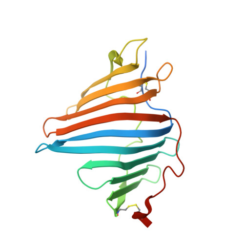
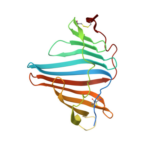
The crystal structure of EPD-BCP1 from a marine sponge


Experimental Data Snapshot

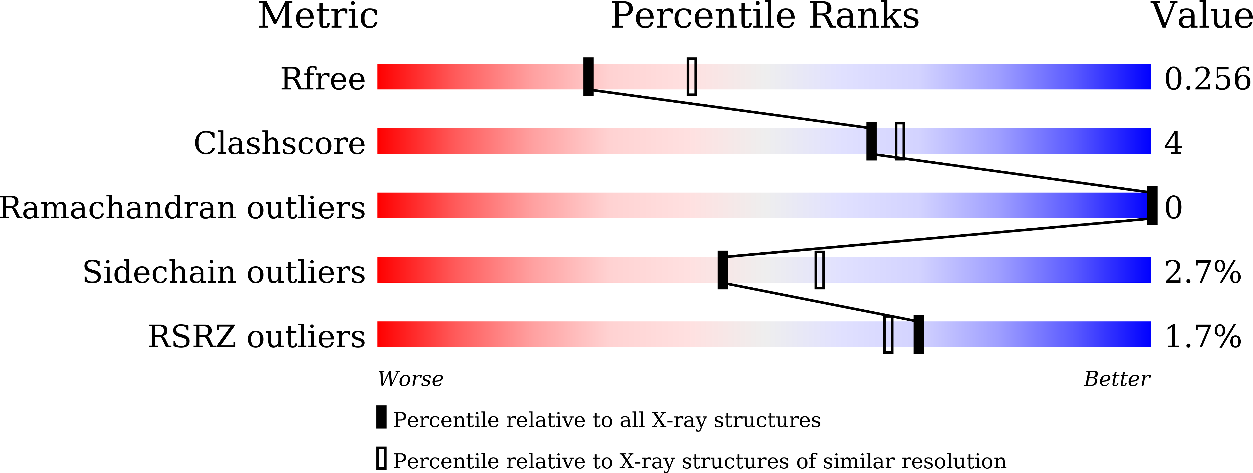
  • Method: X-RAY DIFFRACTION
  • Resolution: 2.44 Å
  • R-Value Free: 0.248 
  • R-Value Work: 0.202 

wwPDB Validation   3D Report Full Report


Ligand Structure Quality Assessment 


This is version 1.1 of the entry. See complete history


Literature

An ependymin-related blue carotenoprotein decorates marine blue sponge.

Kawasaki, S.Kaneko, T.Asano, T.Maoka, T.Takaichi, S.Shomura, Y.

(2023) J Biol Chem 299: 105110-105110

  • DOI: https://doi.org/10.1016/j.jbc.2023.105110
  • Primary Citation of Related Structures:  
    8I34

  • PubMed Abstract: 

    Marine animals display diverse vibrant colors, but the mechanisms underlying their specific coloration remain to be clarified. Blue coloration is known to be achieved through a bathochromic shift of the orange carotenoid astaxanthin (AXT) by the crustacean protein crustacyanin, but other examples have not yet been well investigated. Here, we identified an ependymin (EPD)-related water-soluble blue carotenoprotein responsible for the specific coloration of the marine blue sponge Haliclona sp. EPD was originally identified in the fish brain as a protein involved in memory consolidation and neuronal regeneration. The purified blue protein, designated as EPD-related blue carotenoprotein-1, was identified as a secreted glycoprotein. We show that it consists of a heterodimer that binds orange AXT and mytiloxanthin and exhibits a bathochromic shift. Our crystal structure analysis of the natively purified EPD-related blue carotenoprotein-1 revealed that these two carotenoids are specifically bound to the heterodimer interface, where the polyene chains are aligned in parallel to each other like in β-crustacyanin, although the two proteins are evolutionary and structurally unrelated. Furthermore, using reconstitution assays, we found that incomplete bathochromic shifts occurred when the protein bound to only AXT or mytiloxanthin. Taken together, we identified an EPD in a basal metazoan as a blue protein that decorates the sponge body by binding specific structurally unrelated carotenoids.


  • Organizational Affiliation

    Department of Molecular Microbiology, Tokyo University of Agriculture, Tokyo, Japan. Electronic address: kawashin@nodai.ac.jp.


Macromolecules
Find similar proteins by:  (by identity cutoff)  |  3D Structure
Entity ID: 1
MoleculeChains Sequence LengthOrganismDetailsImage
alpha subunit of EPD-BCP1
A, C, E, G
196Haliclona sp.Mutation(s): 0 
UniProt
Find proteins for A0AA52HX42 (Haliclona sp. kz-2018)
Explore A0AA52HX42 
Go to UniProtKB:  A0AA52HX42
Entity Groups  
Sequence Clusters30% Identity50% Identity70% Identity90% Identity95% Identity100% Identity
UniProt GroupA0AA52HX42
Sequence Annotations
Expand
  • Reference Sequence
Find similar proteins by:  (by identity cutoff)  |  3D Structure
Entity ID: 2
MoleculeChains Sequence LengthOrganismDetailsImage
beta subunit of EPD-BCP1
B, D, F, H
196Haliclona sp.Mutation(s): 0 
UniProt
Find proteins for A0AA52HX50 (Haliclona sp. kz-2018)
Explore A0AA52HX50 
Go to UniProtKB:  A0AA52HX50
Entity Groups  
Sequence Clusters30% Identity50% Identity70% Identity90% Identity95% Identity100% Identity
UniProt GroupA0AA52HX50
Sequence Annotations
Expand
  • Reference Sequence
Oligosaccharides

Help

Entity ID: 3
MoleculeChains Length2D Diagram Glycosylation3D Interactions
2-acetamido-2-deoxy-beta-D-glucopyranose-(1-4)-2-acetamido-2-deoxy-beta-D-glucopyranose
I, J, K, L, M
I, J, K, L, M, N, O, P, Q, R, S, T
2N-Glycosylation
Glycosylation Resources
GlyTouCan:  G42666HT
GlyCosmos:  G42666HT
GlyGen:  G42666HT
Small Molecules
Ligands 2 Unique
IDChains Name / Formula / InChI Key2D Diagram3D Interactions
O1U
Query on O1U

Download Ideal Coordinates CCD File 
AA [auth G],
U [auth A],
W [auth C],
Y [auth E]
(2~{Z},4~{E},6~{E},8~{E},10~{E},12~{E},14~{E},16~{E})-4,8,13,17-tetramethyl-3-oxidanyl-19-[(4~{R})-2,6,6-trimethyl-4-oxidanyl-cyclohexen-1-yl]-1-[(1~{R},4~{S})-1,2,2-trimethyl-4-oxidanyl-cyclopentyl]nonadeca-2,4,6,8,10,12,14,16-octaen-18-yn-1-one
C40 H54 O4
WSLGBPCJDUQFND-BBPSKTQQSA-N
AXT
Query on AXT

Download Ideal Coordinates CCD File 
BA [auth H],
V [auth B],
X [auth D],
Z [auth F]
ASTAXANTHIN
C40 H52 O4
MQZIGYBFDRPAKN-UWFIBFSHSA-N
Experimental Data & Validation

Experimental Data

  • Method: X-RAY DIFFRACTION
  • Resolution: 2.44 Å
  • R-Value Free: 0.248 
  • R-Value Work: 0.202 
  • Space Group: P 1 21 1
Unit Cell:
Length ( Å )Angle ( ˚ )
a = 73.749α = 90
b = 101.38β = 95.06
c = 112.588γ = 90
Software Package:
Software NamePurpose
REFMACrefinement
iMOSFLMdata reduction
Aimlessdata scaling
MOLREPphasing

Structure Validation

View Full Validation Report


Ligand Structure Quality Assessment 


Entry History & Funding Information

Deposition Data


Funding OrganizationLocationGrant Number
Ministry of Education, Culture, Sports, Science and Technology (Japan)JapanS1311017

Revision History  (Full details and data files)

  • Version 1.0: 2023-08-16
    Type: Initial release
  • Version 1.1: 2023-09-06
    Changes: Database references