


Structure of nanobody 11A in complex with coumaphos
Jiadong, L., Hong, W.To be published.
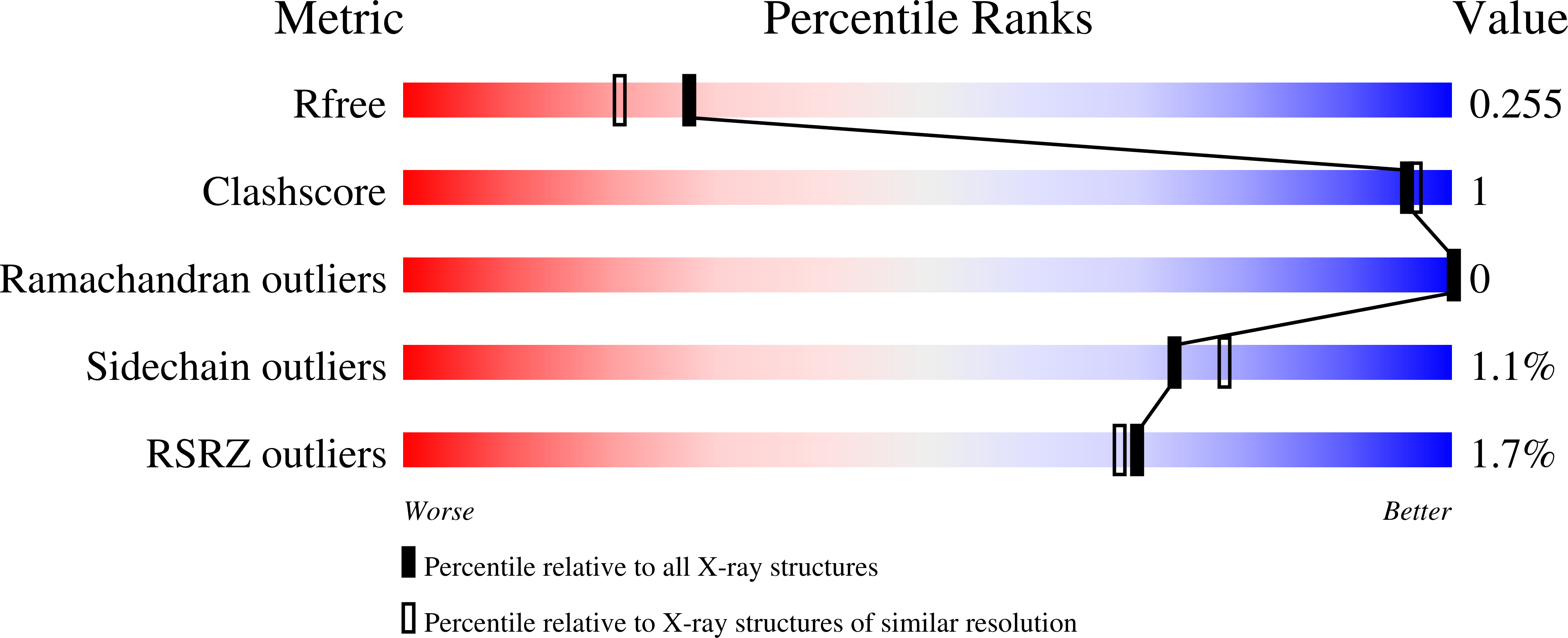
Experimental Data Snapshot
Entity ID: 1 | |||||
|---|---|---|---|---|---|
| Molecule | Chains | Sequence Length | Organism | Details | Image |
| Nanobody 11A | 140 | Camelus bactrianus | Mutation(s): 0 | 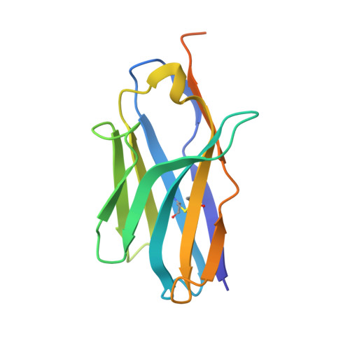 | |
Entity Groups | |||||
| Sequence Clusters | 30% Identity50% Identity70% Identity90% Identity95% Identity100% Identity | ||||
Sequence AnnotationsExpand | |||||
| |||||
| Ligands 1 Unique | |||||
|---|---|---|---|---|---|
| ID | Chains | Name / Formula / InChI Key | 2D Diagram | 3D Interactions | |
| WZ0 (Subject of Investigation/LOI) Query on WZ0 | B [auth A] | coumaphos C14 H16 Cl O5 P S BXNANOICGRISHX-UHFFFAOYSA-N |  | ||
| Length ( Å ) | Angle ( ˚ ) |
|---|---|
| a = 84.166 | α = 90 |
| b = 84.166 | β = 90 |
| c = 32.301 | γ = 120 |
| Software Name | Purpose |
|---|---|
| PHENIX | refinement |
| DIALS | data reduction |
| Aimless | data scaling |
| PHASER | phasing |
| Funding Organization | Location | Grant Number |
|---|---|---|
| Ministry of Science and Technology (MoST, China) | China | 2019YFE0116600 |
RCSB PDB (citation) is hosted by
RCSB PDB is a member of the

