8GK3

Cytochrome P450 3A7 in complex with Dehydroepiandrosterone sulfate

  • Classification: OXIDOREDUCTASE
  • Organism(s): Homo sapiens
  • Expression System: Escherichia coli
  • Mutation(s): No 

  • Deposited: 2023-03-16 Released: 2023-07-26 
  • Deposition Author(s): Liu, J., Scott, E.E.
  • Funding Organization(s): National Institutes of Health/National Institute Of Allergy and Infectious Diseases (NIH/NIAID)

Experimental Data Snapshot

  • Method: X-RAY DIFFRACTION
  • Resolution: 2.60 Å
  • R-Value Free: 0.273 
  • R-Value Work: 0.210 
  • R-Value Observed: 0.210 

wwPDB Validation   3D Report Full Report


Ligand Structure Quality Assessment 


This is version 2.0 of the entry. See complete history


Literature

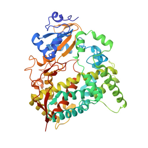
Human cytochrome P450 3A7 binding four copies of its native substrate dehydroepiandrosterone 3-sulfate.

Liu, J.Kandel, S.E.Lampe, J.N.Scott, E.E.

(2023) J Biol Chem 299: 104993-104993

  • DOI: https://doi.org/10.1016/j.jbc.2023.104993
  • Primary Citation of Related Structures:  
    8GK3

  • PubMed Abstract: 

    Human fetal cytochrome P450 3A7 (CYP3A7) is involved in both xenobiotic metabolism and the estriol biosynthetic pathway. Although much is understood about cytochrome P450 3A4 and its role in adult drug metabolism, CYP3A7 is poorly characterized in terms of its interactions with both categories of substrates. Herein, a crystallizable mutated form of CYP3A7 was saturated with its primary endogenous substrate dehydroepiandrosterone 3-sulfate (DHEA-S) to yield a 2.6 Å X-ray structure revealing the unexpected capacity to simultaneously bind four copies of DHEA-S. Two DHEA-S molecules are located in the active site proper, one in a ligand access channel, and one on the hydrophobic F'-G' surface normally embedded in the membrane. While neither DHEA-S binding nor metabolism exhibit cooperative kinetics, the current structure is consistent with cooperativity common to CYP3A enzymes. Overall, this information suggests that mechanism(s) of CYP3A7 interactions with steroidal substrates are complex.


  • Organizational Affiliation

    Department of Medicinal Chemistry, University of Michigan, Ann Arbor, Michigan, USA.


Macromolecules
Find similar proteins by:  (by identity cutoff)  |  3D Structure
Entity ID: 1
MoleculeChains Sequence LengthOrganismDetailsImage
Cytochrome P450 3A7
A, B, C, D, E
A, B, C, D, E, F, G, H, I, J, K, L
486Homo sapiensMutation(s): 0 
Gene Names: CYP3A7
EC: 1.14.14.1
UniProt & NIH Common Fund Data Resources
Find proteins for P24462 (Homo sapiens)
Explore P24462 
Go to UniProtKB:  P24462
PHAROS:  P24462
GTEx:  ENSG00000160870 
Entity Groups  
Sequence Clusters30% Identity50% Identity70% Identity90% Identity95% Identity100% Identity
UniProt GroupP24462
Sequence Annotations
Expand
  • Reference Sequence
Small Molecules
Ligands 2 Unique
IDChains Name / Formula / InChI Key2D Diagram3D Interactions
HEM
Query on HEM

Download Ideal Coordinates CCD File 
DA [auth E]
IA [auth F]
JA [auth G]
KA [auth H]
M [auth A]
DA [auth E],
IA [auth F],
JA [auth G],
KA [auth H],
M [auth A],
OA [auth I],
P [auth B],
RA [auth J],
S [auth C],
UA [auth K],
VA [auth L],
X [auth D]
PROTOPORPHYRIN IX CONTAINING FE
C34 H32 Fe N4 O4
KABFMIBPWCXCRK-RGGAHWMASA-L
ZWY
Query on ZWY

Download Ideal Coordinates CCD File 
AA [auth D]
BA [auth D]
CA [auth D]
EA [auth E]
FA [auth E]
AA [auth D],
BA [auth D],
CA [auth D],
EA [auth E],
FA [auth E],
GA [auth E],
HA [auth E],
LA [auth H],
MA [auth H],
N [auth A],
NA [auth H],
O [auth A],
PA [auth I],
Q [auth B],
QA [auth I],
R [auth B],
SA [auth J],
T [auth C],
TA [auth J],
U [auth C],
V [auth C],
W [auth C],
Y [auth D],
Z [auth D]
17-oxoandrost-5-en-3beta-yl hydrogen sulfate
C19 H28 O5 S
CZWCKYRVOZZJNM-USOAJAOKSA-N
Experimental Data & Validation

Experimental Data

  • Method: X-RAY DIFFRACTION
  • Resolution: 2.60 Å
  • R-Value Free: 0.273 
  • R-Value Work: 0.210 
  • R-Value Observed: 0.210 
  • Space Group: P 1 21 1
Unit Cell:
Length ( Å )Angle ( ˚ )
a = 114.516α = 90
b = 219.381β = 102.165
c = 130.447γ = 90
Software Package:
Software NamePurpose
PHENIXrefinement
PHASERphasing
Cootmodel building
SCALAdata scaling
Blu-Icedata collection
XDSdata reduction

Structure Validation

View Full Validation Report


Ligand Structure Quality Assessment 


Entry History & Funding Information

Deposition Data


Funding OrganizationLocationGrant Number
National Institutes of Health/National Institute Of Allergy and Infectious Diseases (NIH/NIAID)United StatesRO1 AI150494

Revision History  (Full details and data files)

  • Version 1.0: 2023-07-26
    Type: Initial release
  • Version 1.1: 2023-08-09
    Changes: Database references
  • Version 2.0: 2023-08-30
    Type: Coordinate replacement
    Reason: Ligand geometry
    Changes: Advisory, Atomic model, Author supporting evidence, Data collection, Derived calculations, Non-polymer description, Refinement description, Structure summary