8FOI

Native GABA-A receptor from the mouse brain, alpha1-beta2-gamma2 subtype, in complex with GABA and allopregnanolone

  • Classification: MEMBRANE PROTEIN
  • Organism(s): Mus musculus
  • Expression System: Spodoptera frugiperda
  • Mutation(s): No 

  • Deposited: 2022-12-30 Released: 2023-09-20 
  • Deposition Author(s): Sun, C., Gouaux, E.
  • Funding Organization(s): National Institutes of Health/National Institute of General Medical Sciences (NIH/NIGMS), Howard Hughes Medical Institute (HHMI)

Experimental Data Snapshot

  • Method: ELECTRON MICROSCOPY
  • Resolution: 2.50 Å
  • Aggregation State: PARTICLE 
  • Reconstruction Method: SINGLE PARTICLE 

wwPDB Validation   3D Report Full Report


This is version 2.0 of the entry. See complete history


Literature

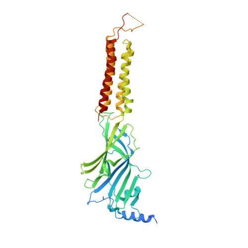
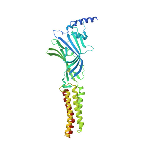
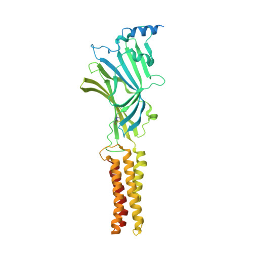
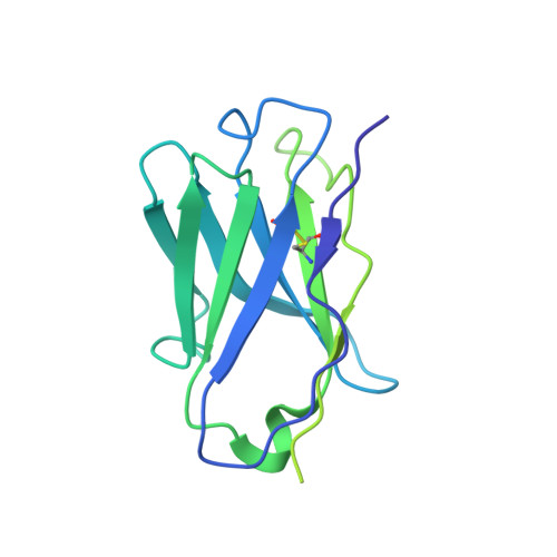
Cryo-EM structures reveal native GABA A receptor assemblies and pharmacology.

Sun, C.Zhu, H.Clark, S.Gouaux, E.

(2023) Nature 622: 195-201

  • DOI: https://doi.org/10.1038/s41586-023-06556-w
  • Primary Citation of Related Structures:  
    8FOI, 8G4N, 8G4O, 8G4X, 8G5F, 8G5G, 8G5H

  • PubMed Abstract: 

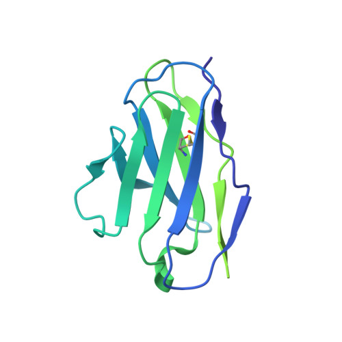
    Type A γ-aminobutyric acid receptors (GABA A Rs) are the principal inhibitory receptors in the brain and the target of a wide range of clinical agents, including anaesthetics, sedatives, hypnotics and antidepressants 1-3 . However, our understanding of GABA A R pharmacology has been hindered by the vast number of pentameric assemblies that can be derived from 19 different subunits 4 and the lack of structural knowledge of clinically relevant receptors. Here, we isolate native murine GABA A R assemblies containing the widely expressed α1 subunit and elucidate their structures in complex with drugs used to treat insomnia (zolpidem (ZOL) and flurazepam) and postpartum depression (the neurosteroid allopregnanolone (APG)). Using cryo-electron microscopy (cryo-EM) analysis and single-molecule photobleaching experiments, we uncover three major structural populations in the brain: the canonical α1β2γ2 receptor containing two α1 subunits, and two assemblies containing one α1 and either an α2 or α3 subunit, in which the single α1-containing receptors feature a more compact arrangement between the transmembrane and extracellular domains. Interestingly, APG is bound at the transmembrane α/β subunit interface, even when not added to the sample, revealing an important role for endogenous neurosteroids in modulating native GABA A Rs. Together with structurally engaged lipids, neurosteroids produce global conformational changes throughout the receptor that modify the ion channel pore and the binding sites for GABA and insomnia medications. Our data reveal the major α1-containing GABA A R assemblies, bound with endogenous neurosteroid, thus defining a structural landscape from which subtype-specific drugs can be developed.


  • Organizational Affiliation

    Vollum Institute, Oregon Health and Science University, Portland, OR, USA.


Macromolecules
Find similar proteins by:  (by identity cutoff)  |  3D Structure
Entity ID: 1
MoleculeChains Sequence LengthOrganismDetailsImage
Gamma-aminobutyric acid receptor subunit alpha-1
A, C
455Mus musculusMutation(s): 0 
UniProt
Find proteins for P62812 (Mus musculus)
Explore P62812 
Go to UniProtKB:  P62812
Entity Groups  
Sequence Clusters30% Identity50% Identity70% Identity90% Identity95% Identity100% Identity
UniProt GroupP62812
Sequence Annotations
Expand
  • Reference Sequence
Find similar proteins by:  (by identity cutoff)  |  3D Structure
Entity ID: 2
MoleculeChains Sequence LengthOrganismDetailsImage
Gamma-aminobutyric acid receptor subunit beta-2
B, E
512Mus musculusMutation(s): 0 
UniProt
Find proteins for P63137 (Mus musculus)
Explore P63137 
Go to UniProtKB:  P63137
Entity Groups  
Sequence Clusters30% Identity50% Identity70% Identity90% Identity95% Identity100% Identity
UniProt GroupP63137
Sequence Annotations
Expand
  • Reference Sequence
Find similar proteins by:  (by identity cutoff)  |  3D Structure
Entity ID: 3
MoleculeChains Sequence LengthOrganismDetailsImage
Gamma-aminobutyric acid receptor subunit gamma-2474Mus musculusMutation(s): 0 
UniProt & NIH Common Fund Data Resources
Find proteins for P22723 (Mus musculus)
Explore P22723 
Go to UniProtKB:  P22723
IMPC:  MGI:95623
Entity Groups  
Sequence Clusters30% Identity50% Identity70% Identity90% Identity95% Identity100% Identity
UniProt GroupP22723
Sequence Annotations
Expand
  • Reference Sequence
Find similar proteins by:  (by identity cutoff)  |  3D Structure
Entity ID: 4
MoleculeChains Sequence LengthOrganismDetailsImage
Heavy Chain of 8E3 FabF [auth H],
G [auth J]
223Mus musculusMutation(s): 0 
Entity Groups  
Sequence Clusters30% Identity50% Identity70% Identity90% Identity95% Identity100% Identity
Sequence Annotations
Expand
  • Reference Sequence
Find similar proteins by:  (by identity cutoff)  |  3D Structure
Entity ID: 5
MoleculeChains Sequence LengthOrganismDetailsImage
Light Chain of 8E3-FabH [auth K],
I [auth L]
213Mus musculusMutation(s): 0 
Entity Groups  
Sequence Clusters30% Identity50% Identity70% Identity90% Identity95% Identity100% Identity
Sequence Annotations
Expand
  • Reference Sequence
Oligosaccharides

Help

Entity ID: 6
MoleculeChains Length2D Diagram Glycosylation3D Interactions
alpha-D-mannopyranose-(1-2)-alpha-D-mannopyranose-(1-3)-[alpha-D-mannopyranose-(1-3)-alpha-D-mannopyranose-(1-6)]beta-D-mannopyranose-(1-4)-2-acetamido-2-deoxy-beta-D-glucopyranose-(1-4)-2-acetamido-2-deoxy-beta-D-glucopyranoseJ [auth F]7N-Glycosylation
Glycosylation Resources
GlyTouCan:  G07617FP
GlyCosmos:  G07617FP
GlyGen:  G07617FP
Entity ID: 7
MoleculeChains Length2D Diagram Glycosylation3D Interactions
alpha-D-mannopyranose-(1-3)-[alpha-D-mannopyranose-(1-6)]beta-D-mannopyranose-(1-4)-2-acetamido-2-deoxy-beta-D-glucopyranose-(1-4)-2-acetamido-2-deoxy-beta-D-glucopyranoseK [auth G],
L [auth I],
M,
O,
P
5N-Glycosylation
Glycosylation Resources
GlyTouCan:  G22768VO
GlyCosmos:  G22768VO
GlyGen:  G22768VO
Entity ID: 8
MoleculeChains Length2D Diagram Glycosylation3D Interactions
beta-D-mannopyranose-(1-4)-2-acetamido-2-deoxy-beta-D-glucopyranose-(1-4)-2-acetamido-2-deoxy-beta-D-glucopyranose
N
3N-Glycosylation
Glycosylation Resources
GlyTouCan:  G15407YE
GlyCosmos:  G15407YE
GlyGen:  G15407YE
Small Molecules
Ligands 7 Unique
IDChains Name / Formula / InChI Key2D Diagram3D Interactions
POV
Query on POV

Download Ideal Coordinates CCD File 
IB [auth E],
OA [auth C],
PA [auth C],
X [auth A],
Y [auth A]
(2S)-3-(hexadecanoyloxy)-2-[(9Z)-octadec-9-enoyloxy]propyl 2-(trimethylammonio)ethyl phosphate
C42 H82 N O8 P
WTJKGGKOPKCXLL-PFDVCBLKSA-N
PIO
Query on PIO

Download Ideal Coordinates CCD File 
IA [auth C],
Q [auth A]
[(2R)-2-octanoyloxy-3-[oxidanyl-[(1R,2R,3S,4R,5R,6S)-2,3,6-tris(oxidanyl)-4,5-diphosphonooxy-cyclohexyl]oxy-phosphoryl]oxy-propyl] octanoate
C25 H49 O19 P3
XLNCEHRXXWQMPK-MJUMVPIBSA-N
Y4B (Subject of Investigation/LOI)
Query on Y4B

Download Ideal Coordinates CCD File 
AA [auth B],
YA [auth E]
allopregnanolone
C21 H34 O2
AURFZBICLPNKBZ-SYBPFIFISA-N
NAG
Query on NAG

Download Ideal Coordinates CCD File 
QA [auth D]2-acetamido-2-deoxy-beta-D-glucopyranose
C8 H15 N O6
OVRNDRQMDRJTHS-FMDGEEDCSA-N
D12
Query on D12

Download Ideal Coordinates CCD File 
AB [auth E]
BA [auth B]
CA [auth B]
CB [auth E]
DA [auth B]
AB [auth E],
BA [auth B],
CA [auth B],
CB [auth E],
DA [auth B],
EA [auth B],
FA [auth B],
GA [auth B],
GB [auth E],
HA [auth B],
JB [auth E],
MA [auth C],
NA [auth C],
R [auth A],
S [auth A],
VA [auth D],
WA [auth D]
DODECANE
C12 H26
SNRUBQQJIBEYMU-UHFFFAOYSA-N
OCT
Query on OCT

Download Ideal Coordinates CCD File 
BB [auth E]
DB [auth E]
EB [auth E]
FB [auth E]
HB [auth E]
BB [auth E],
DB [auth E],
EB [auth E],
FB [auth E],
HB [auth E],
JA [auth C],
KA [auth C],
LA [auth C],
RA [auth D],
SA [auth D],
T [auth A],
TA [auth D],
U [auth A],
UA [auth D],
V [auth A],
W [auth A],
XA [auth D]
N-OCTANE
C8 H18
TVMXDCGIABBOFY-UHFFFAOYSA-N
ABU (Subject of Investigation/LOI)
Query on ABU

Download Ideal Coordinates CCD File 
Z [auth B],
ZA [auth E]
GAMMA-AMINO-BUTANOIC ACID
C4 H9 N O2
BTCSSZJGUNDROE-UHFFFAOYSA-N
Experimental Data & Validation

Experimental Data

  • Method: ELECTRON MICROSCOPY
  • Resolution: 2.50 Å
  • Aggregation State: PARTICLE 
  • Reconstruction Method: SINGLE PARTICLE 
EM Software:
TaskSoftware PackageVersion
RECONSTRUCTIONcryoSPARC3.3.2

Structure Validation

View Full Validation Report


Entry History & Funding Information

Deposition Data


Funding OrganizationLocationGrant Number
National Institutes of Health/National Institute of General Medical Sciences (NIH/NIGMS)United States5R01GM100400-08
Howard Hughes Medical Institute (HHMI)United States--

Revision History  (Full details and data files)

  • Version 1.0: 2023-09-20
    Type: Initial release
  • Version 1.1: 2023-10-04
    Changes: Database references
  • Version 1.2: 2023-10-18
    Changes: Database references
  • Version 2.0: 2023-11-15
    Changes: Advisory, Atomic model, Data collection