8FHS

Human L-type voltage-gated calcium channel Cav1.2 in the presence of amiodarone and sofosbuvir at 3.3 Angstrom resolution


Experimental Data Snapshot

  • Method: ELECTRON MICROSCOPY
  • Resolution: 3.30 Å
  • Aggregation State: PARTICLE 
  • Reconstruction Method: SINGLE PARTICLE 

wwPDB Validation   3D Report Full Report


This is version 1.0 of the entry. See complete history


Literature

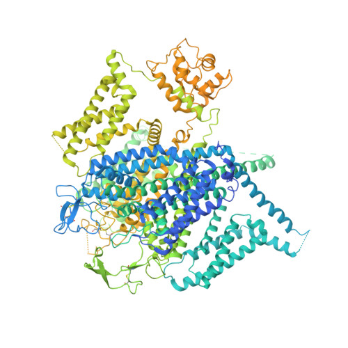
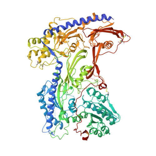
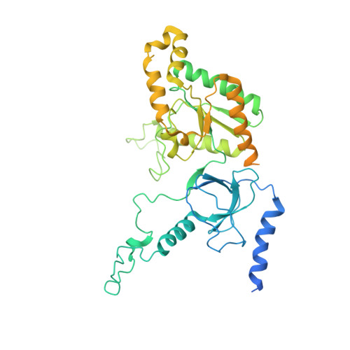
Structural basis for human Ca v 1.2 inhibition by multiple drugs and the neurotoxin calciseptine.

Gao, S.Yao, X.Chen, J.Huang, G.Fan, X.Xue, L.Li, Z.Wu, T.Zheng, Y.Huang, J.Jin, X.Wang, Y.Wang, Z.Yu, Y.Liu, L.Pan, X.Song, C.Yan, N.

(2023) Cell 186: 5363-5374.e16

  • DOI: https://doi.org/10.1016/j.cell.2023.10.007
  • Primary Citation of Related Structures:  
    8FHS, 8WE6, 8WE7, 8WE8, 8WE9, 8WEA

  • PubMed Abstract: 

    Ca v 1.2 channels play crucial roles in various neuronal and physiological processes. Here, we present cryo-EM structures of human Ca v 1.2, both in its apo form and in complex with several drugs, as well as the peptide neurotoxin calciseptine. Most structures, apo or bound to calciseptine, amlodipine, or a combination of amiodarone and sofosbuvir, exhibit a consistent inactivated conformation with a sealed gate, three up voltage-sensing domains (VSDs), and a down VSD II . Calciseptine sits on the shoulder of the pore domain, away from the permeation path. In contrast, when pinaverium bromide, an antispasmodic drug, is inserted into a cavity reminiscent of the IFM-binding site in Na v channels, a series of structural changes occur, including upward movement of VSD II coupled with dilation of the selectivity filter and its surrounding segments in repeat III. Meanwhile, S4-5 III merges with S5 III to become a single helix, resulting in a widened but still non-conductive intracellular gate.


  • Organizational Affiliation

    Department of Urology, Zhongnan Hospital of Wuhan University, TaiKang Center for Life and Medical Sciences, School of Pharmaceutical Sciences, Wuhan University, Wuhan 430071, China; Department of Molecular Biology, Princeton University, Princeton, NJ 08544, USA. Electronic address: gaoshuai812@whu.edu.cn.


Macromolecules
Find similar proteins by:  (by identity cutoff)  |  3D Structure
Entity ID: 1
MoleculeChains Sequence LengthOrganismDetailsImage
Voltage-dependent L-type calcium channel subunit alpha-1C2,221Homo sapiensMutation(s): 0 
Gene Names: CACNA1CCACH2CACN2CACNL1A1CCHL1A1
Membrane Entity: Yes 
UniProt & NIH Common Fund Data Resources
Find proteins for Q13936 (Homo sapiens)
Explore Q13936 
Go to UniProtKB:  Q13936
PHAROS:  Q13936
GTEx:  ENSG00000151067 
Entity Groups  
Sequence Clusters30% Identity50% Identity70% Identity90% Identity95% Identity100% Identity
UniProt GroupQ13936
Sequence Annotations
Expand
  • Reference Sequence
Find similar proteins by:  (by identity cutoff)  |  3D Structure
Entity ID: 2
MoleculeChains Sequence LengthOrganismDetailsImage
Voltage-dependent calcium channel subunit alpha-2/delta-1B [auth D]1,103Homo sapiensMutation(s): 0 
Gene Names: CACNA2D1CACNL2ACCHL2AMHS3
Membrane Entity: Yes 
UniProt & NIH Common Fund Data Resources
Find proteins for P54289 (Homo sapiens)
Explore P54289 
Go to UniProtKB:  P54289
PHAROS:  P54289
GTEx:  ENSG00000153956 
Entity Groups  
Sequence Clusters30% Identity50% Identity70% Identity90% Identity95% Identity100% Identity
UniProt GroupP54289
Sequence Annotations
Expand
  • Reference Sequence
Find similar proteins by:  (by identity cutoff)  |  3D Structure
Entity ID: 3
MoleculeChains Sequence LengthOrganismDetailsImage
Voltage-dependent L-type calcium channel subunit beta-3484Homo sapiensMutation(s): 0 
Gene Names: CACNB3CACNLB3
UniProt & NIH Common Fund Data Resources
Find proteins for P54284 (Homo sapiens)
Explore P54284 
Go to UniProtKB:  P54284
PHAROS:  P54284
GTEx:  ENSG00000167535 
Entity Groups  
Sequence Clusters30% Identity50% Identity70% Identity90% Identity95% Identity100% Identity
UniProt GroupP54284
Sequence Annotations
Expand
  • Reference Sequence
Oligosaccharides

Help

Entity ID: 4
MoleculeChains Length2D Diagram Glycosylation3D Interactions
2-acetamido-2-deoxy-beta-D-glucopyranose-(1-4)-2-acetamido-2-deoxy-beta-D-glucopyranose-(1-4)-2-acetamido-2-deoxy-beta-D-glucopyranoseD [auth B]3N-Glycosylation
Glycosylation Resources
GlyTouCan:  G47362BJ
GlyCosmos:  G47362BJ
GlyGen:  G47362BJ
Entity ID: 5
MoleculeChains Length2D Diagram Glycosylation3D Interactions
2-acetamido-2-deoxy-beta-D-glucopyranose-(1-4)-2-acetamido-2-deoxy-beta-D-glucopyranose
E, G, H
2N-Glycosylation
Glycosylation Resources
GlyTouCan:  G42666HT
GlyCosmos:  G42666HT
GlyGen:  G42666HT
Entity ID: 6
MoleculeChains Length2D Diagram Glycosylation3D Interactions
2-acetamido-2-deoxy-beta-D-glucopyranose-(1-4)-2-acetamido-2-deoxy-beta-D-glucopyranose-(1-4)-2-acetamido-2-deoxy-beta-D-glucopyranose-(1-4)-2-acetamido-2-deoxy-beta-D-glucopyranose
F
4N-Glycosylation
Glycosylation Resources
GlyTouCan:  G01361SX
GlyCosmos:  G01361SX
GlyGen:  G01361SX
Small Molecules
Ligands 6 Unique
IDChains Name / Formula / InChI Key2D Diagram3D Interactions
3PE
Query on 3PE

Download Ideal Coordinates CCD File 
L [auth A],
N [auth A]
1,2-Distearoyl-sn-glycerophosphoethanolamine
C41 H82 N O8 P
LVNGJLRDBYCPGB-LDLOPFEMSA-N
BBI (Subject of Investigation/LOI)
Query on BBI

Download Ideal Coordinates CCD File 
J [auth A](2-butyl-1-benzofuran-3-yl){4-[2-(diethylamino)ethoxy]-3,5-diiodophenyl}methanone
C25 H29 I2 N O3
IYIKLHRQXLHMJQ-UHFFFAOYSA-N
WG6 (Subject of Investigation/LOI)
Query on WG6

Download Ideal Coordinates CCD File 
K [auth A]Sofosbuvir
C22 H29 F N3 O9 P
TTZHDVOVKQGIBA-IQWMDFIBSA-N
CLR
Query on CLR

Download Ideal Coordinates CCD File 
M [auth A],
O [auth A],
P [auth A],
Q [auth A]
CHOLESTEROL
C27 H46 O
HVYWMOMLDIMFJA-DPAQBDIFSA-N
NAG
Query on NAG

Download Ideal Coordinates CCD File 
S [auth D],
T [auth D]
2-acetamido-2-deoxy-beta-D-glucopyranose
C8 H15 N O6
OVRNDRQMDRJTHS-FMDGEEDCSA-N
CA
Query on CA

Download Ideal Coordinates CCD File 
I [auth A],
R [auth D]
CALCIUM ION
Ca
BHPQYMZQTOCNFJ-UHFFFAOYSA-N
Experimental Data & Validation

Experimental Data

  • Method: ELECTRON MICROSCOPY
  • Resolution: 3.30 Å
  • Aggregation State: PARTICLE 
  • Reconstruction Method: SINGLE PARTICLE 

Structure Validation

View Full Validation Report


Entry History & Funding Information

Deposition Data


Funding OrganizationLocationGrant Number
Other privateChinaZ191100001119127

Revision History  (Full details and data files)

  • Version 1.0: 2023-12-13
    Type: Initial release