8FD7

Structure of the human L-type voltage-gated calcium channel Cav1.2 complexed with gabapentin


Experimental Data Snapshot

  • Method: ELECTRON MICROSCOPY
  • Resolution: 3.10 Å
  • Aggregation State: PARTICLE 
  • Reconstruction Method: SINGLE PARTICLE 

wwPDB Validation   3D Report Full Report


This is version 1.2 of the entry. See complete history


Literature

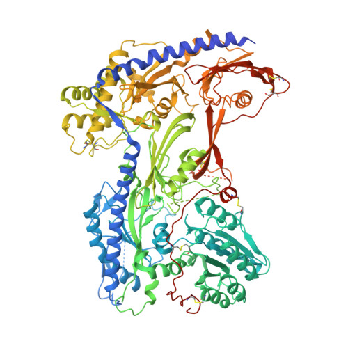
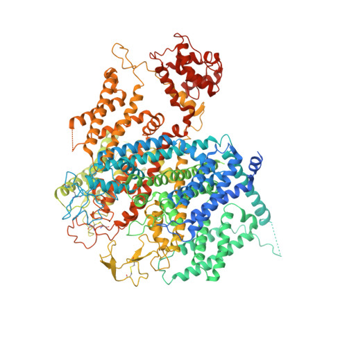
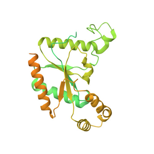
Structural basis for Ca V alpha 2 delta :gabapentin binding.

Chen, Z.Mondal, A.Minor Jr., D.L.

(2023) Nat Struct Mol Biol 30: 735-739

  • DOI: https://doi.org/10.1038/s41594-023-00951-7
  • Primary Citation of Related Structures:  
    8FD7

  • PubMed Abstract: 

    Gabapentinoid drugs for pain and anxiety act on the Ca V α 2 δ-1 and Ca V α 2 δ-2 subunits of high-voltage-activated calcium channels (Ca V 1s and Ca V 2s). Here we present the cryo-EM structure of the gabapentin-bound brain and cardiac Ca V 1.2/Ca V β 3 /Ca V α 2 δ-1 channel. The data reveal a binding pocket in the Ca V α 2 δ-1 dCache1 domain that completely encapsulates gabapentin and define Ca V α 2 δ isoform sequence variations that explain the gabapentin binding selectivity of Ca V α 2 δ-1 and Ca V α 2 δ-2.


  • Organizational Affiliation

    Cardiovascular Research Institute, University of California, San Francisco, CA, USA.


Macromolecules
Find similar proteins by:  (by identity cutoff)  |  3D Structure
Entity ID: 1
MoleculeChains Sequence LengthOrganismDetailsImage
Voltage-dependent calcium channel subunit alpha-2/delta-1A [auth D]1,105Oryctolagus cuniculusMutation(s): 0 
Gene Names: CACNA2D1CACNL2ACCHL2A
Membrane Entity: Yes 
UniProt
Find proteins for P13806 (Oryctolagus cuniculus)
Explore P13806 
Go to UniProtKB:  P13806
Entity Groups  
Sequence Clusters30% Identity50% Identity70% Identity90% Identity95% Identity100% Identity
UniProt GroupP13806
Sequence Annotations
Expand
  • Reference Sequence
Find similar proteins by:  (by identity cutoff)  |  3D Structure
Entity ID: 2
MoleculeChains Sequence LengthOrganismDetailsImage
Voltage-dependent L-type calcium channel subunit alpha-1CB [auth K]1,648Homo sapiensMutation(s): 0 
Gene Names: CACNA1CCACH2CACN2CACNL1A1CCHL1A1
Membrane Entity: Yes 
UniProt & NIH Common Fund Data Resources
Find proteins for Q13936 (Homo sapiens)
Explore Q13936 
Go to UniProtKB:  Q13936
PHAROS:  Q13936
GTEx:  ENSG00000151067 
Entity Groups  
Sequence Clusters30% Identity50% Identity70% Identity90% Identity95% Identity100% Identity
UniProt GroupQ13936
Sequence Annotations
Expand
  • Reference Sequence
Find similar proteins by:  (by identity cutoff)  |  3D Structure
Entity ID: 3
MoleculeChains Sequence LengthOrganismDetailsImage
Voltage-dependent L-type calcium channel subunit beta-3477Oryctolagus cuniculusMutation(s): 0 
Gene Names: CACNB3CACNLB3
UniProt
Find proteins for P54286 (Oryctolagus cuniculus)
Explore P54286 
Go to UniProtKB:  P54286
Entity Groups  
Sequence Clusters30% Identity50% Identity70% Identity90% Identity95% Identity100% Identity
UniProt GroupP54286
Sequence Annotations
Expand
  • Reference Sequence
Oligosaccharides

Help

Entity ID: 4
MoleculeChains Length2D Diagram Glycosylation3D Interactions
2-acetamido-2-deoxy-beta-D-glucopyranose-(1-4)-2-acetamido-2-deoxy-beta-D-glucopyranoseD [auth F]2N/A
Glycosylation Resources
GlyTouCan:  G42666HT
GlyCosmos:  G42666HT
GlyGen:  G42666HT
Small Molecules
Ligands 7 Unique
IDChains Name / Formula / InChI Key2D Diagram3D Interactions
WO9
Query on WO9

Download Ideal Coordinates CCD File 
P [auth K](2R)-3-{[(R)-(2-aminoethoxy)(hydroxy)phosphoryl]oxy}-2-(dodecanoyloxy)propyl heptadecanoate
C34 H68 N O8 P
YIIGGWPZRIXGNH-JGCGQSQUSA-N
YSW
Query on YSW

Download Ideal Coordinates CCD File 
Q [auth K](2S)-3-{[(R)-(2-aminoethoxy)(hydroxy)phosphoryl]oxy}-2-(dodecanoyloxy)propyl dodecanoate
C29 H58 N O8 P
ZLGYVWRJIZPQMM-MHZLTWQESA-N
CLR
Query on CLR

Download Ideal Coordinates CCD File 
R [auth K],
S [auth K],
T [auth K]
CHOLESTEROL
C27 H46 O
HVYWMOMLDIMFJA-DPAQBDIFSA-N
NAG
Query on NAG

Download Ideal Coordinates CCD File 
E [auth D]
G [auth D]
H [auth D]
I [auth D]
J [auth D]
E [auth D],
G [auth D],
H [auth D],
I [auth D],
J [auth D],
K [auth D],
L [auth D],
M [auth D]
2-acetamido-2-deoxy-beta-D-glucopyranose
C8 H15 N O6
OVRNDRQMDRJTHS-FMDGEEDCSA-N
GBN
Query on GBN

Download Ideal Coordinates CCD File 
F [auth D][1-(AMINOMETHYL)CYCLOHEXYL]ACETIC ACID
C9 H17 N O2
UGJMXCAKCUNAIE-UHFFFAOYSA-N
CA
Query on CA

Download Ideal Coordinates CCD File 
N [auth D],
U [auth K],
V [auth K]
CALCIUM ION
Ca
BHPQYMZQTOCNFJ-UHFFFAOYSA-N
NA
Query on NA

Download Ideal Coordinates CCD File 
O [auth D]SODIUM ION
Na
FKNQFGJONOIPTF-UHFFFAOYSA-N
Experimental Data & Validation

Experimental Data

  • Method: ELECTRON MICROSCOPY
  • Resolution: 3.10 Å
  • Aggregation State: PARTICLE 
  • Reconstruction Method: SINGLE PARTICLE 

Structure Validation

View Full Validation Report


Entry History & Funding Information

Deposition Data


Funding OrganizationLocationGrant Number
National Institutes of Health/National Heart, Lung, and Blood Institute (NIH/NHLBI)United StatesHL080050
National Institutes of Health/National Institute on Deafness and Other Communication Disorders (NIH/NIDCD)United StatesDC007664

Revision History  (Full details and data files)

  • Version 1.0: 2023-03-22
    Type: Initial release
  • Version 1.1: 2023-04-12
    Changes: Database references
  • Version 1.2: 2023-07-05
    Changes: Database references