8ERP

Structure of Xenopus cholinephosphotransferase1 in complex with CDP-choline

  • Classification: TRANSFERASE
  • Organism(s): Xenopus laevis
  • Expression System: Spodoptera frugiperda
  • Mutation(s): No 
  • Membrane Protein: Yes  OPMPDBTMmpstruc

  • Deposited: 2022-10-12 Released: 2023-04-26 
  • Deposition Author(s): Wang, L., Zhou, M.
  • Funding Organization(s): National Institutes of Health/National Institute of Diabetes and Digestive and Kidney Disease (NIH/NIDDK), National Institutes of Health/National Heart, Lung, and Blood Institute (NIH/NHLBI), Cancer Prevention and Research Institute of Texas (CPRIT)

Experimental Data Snapshot

  • Method: ELECTRON MICROSCOPY
  • Resolution: 3.70 Å
  • Aggregation State: PARTICLE 
  • Reconstruction Method: SINGLE PARTICLE 

wwPDB Validation   3D Report Full Report


This is version 1.2 of the entry. See complete history


Literature

Structure of a eukaryotic cholinephosphotransferase-1 reveals mechanisms of substrate recognition and catalysis.

Wang, L.Zhou, M.

(2023) Nat Commun 14: 2753-2753

  • DOI: https://doi.org/10.1038/s41467-023-38003-9
  • Primary Citation of Related Structures:  
    8ERO, 8ERP

  • PubMed Abstract: 

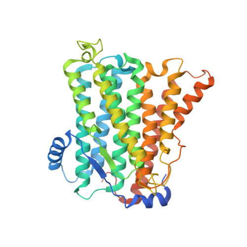
    Phosphatidylcholine (PC) is the most abundant phospholipid in eukaryotic cell membranes. In eukaryotes, two highly homologous enzymes, cholinephosphotransferase-1 (CHPT1) and choline/ethanolamine phosphotransferase-1 (CEPT1) catalyze the final step of de novo PC synthesis. CHPT1/CEPT1 joins two substrates, cytidine diphosphate-choline (CDP-choline) and diacylglycerol (DAG), to produce PC, and Mg 2+ is required for the reaction. However, mechanisms of substrate recognition and catalysis remain unresolved. Here we report structures of a CHPT1 from Xenopus laevis (xlCHPT1) determined by cryo-electron microscopy to an overall resolution of ~3.2 Å. xlCHPT1 forms a homodimer, and each protomer has 10 transmembrane helices (TMs). The first 6 TMs carve out a cone-shaped enclosure in the membrane in which the catalysis occurs. The enclosure opens to the cytosolic side, where a CDP-choline and two Mg 2+ are coordinated. The structures identify a catalytic site unique to eukaryotic CHPT1/CEPT1 and suggest an entryway for DAG. The structures also reveal an internal pseudo two-fold symmetry between TM3-6 and TM7-10, and suggest that CHPT1/CEPT1 may have evolved from their distant prokaryotic ancestors through gene duplication.


  • Organizational Affiliation

    Verna and Marrs McLean Department of Biochemistry and Molecular Biology, Baylor College of Medicine, Houston, TX, USA. liew@bcm.edu.


Macromolecules
Find similar proteins by:  (by identity cutoff)  |  3D Structure
Entity ID: 1
MoleculeChains Sequence LengthOrganismDetailsImage
Cholinephosphotransferase 1A [auth B],
B [auth A]
402Xenopus laevisMutation(s): 0 
Gene Names: chpt1
EC: 2.7.8.2
Membrane Entity: Yes 
UniProt
Find proteins for Q4KLV1 (Xenopus laevis)
Explore Q4KLV1 
Go to UniProtKB:  Q4KLV1
Entity Groups  
Sequence Clusters30% Identity50% Identity70% Identity90% Identity95% Identity100% Identity
UniProt GroupQ4KLV1
Sequence Annotations
Expand
  • Reference Sequence
Small Molecules
Ligands 3 Unique
IDChains Name / Formula / InChI Key2D Diagram3D Interactions
LBN
Query on LBN

Download Ideal Coordinates CCD File 
AA [auth A]
BA [auth A]
E [auth B]
F [auth B]
H [auth B]
AA [auth A],
BA [auth A],
E [auth B],
F [auth B],
H [auth B],
I [auth B],
J [auth B],
K [auth B],
L [auth B],
M [auth B],
N [auth B],
O [auth B],
P [auth A],
Q [auth A],
T [auth A],
U [auth A],
W [auth A],
X [auth A],
Y [auth A],
Z [auth A]
1-palmitoyl-2-oleoyl-sn-glycero-3-phosphocholine
C42 H82 N O8 P
WTJKGGKOPKCXLL-VYOBOKEXSA-N
CDC (Subject of Investigation/LOI)
Query on CDC

Download Ideal Coordinates CCD File 
G [auth B],
V [auth A]
[2-CYTIDYLATE-O'-PHOSPHONYLOXYL]-ETHYL-TRIMETHYL-AMMONIUM
C14 H26 N4 O11 P2
RZZPDXZPRHQOCG-OJAKKHQRSA-N
MG
Query on MG

Download Ideal Coordinates CCD File 
C [auth B],
D [auth B],
R [auth A],
S [auth A]
MAGNESIUM ION
Mg
JLVVSXFLKOJNIY-UHFFFAOYSA-N
Experimental Data & Validation

Experimental Data

  • Method: ELECTRON MICROSCOPY
  • Resolution: 3.70 Å
  • Aggregation State: PARTICLE 
  • Reconstruction Method: SINGLE PARTICLE 

Structure Validation

View Full Validation Report


Entry History & Funding Information

Deposition Data


Funding OrganizationLocationGrant Number
National Institutes of Health/National Institute of Diabetes and Digestive and Kidney Disease (NIH/NIDDK)United StatesDK122784
National Institutes of Health/National Heart, Lung, and Blood Institute (NIH/NHLBI)United StatesHL086392
Cancer Prevention and Research Institute of Texas (CPRIT)United StatesR1223

Revision History  (Full details and data files)

  • Version 1.0: 2023-04-26
    Type: Initial release
  • Version 1.1: 2023-05-03
    Changes: Database references, Structure summary
  • Version 1.2: 2023-11-08
    Changes: Data collection, Database references