8CXW

CamA Adenine Methyltransferase Complexed to Cognate Substrate DNA and Inhibitor piclidenoson (Compound 4)


Experimental Data Snapshot

  • Method: X-RAY DIFFRACTION
  • Resolution: 2.78 Å
  • R-Value Free: 0.227 
  • R-Value Work: 0.184 
  • R-Value Observed: 0.186 

wwPDB Validation   3D Report Full Report


Ligand Structure Quality Assessment 


This is version 1.3 of the entry. See complete history


Literature

Systematic Design of Adenosine Analogs as Inhibitors of a Clostridioides difficile- Specific DNA Adenine Methyltransferase Required for Normal Sporulation and Persistence.

Zhou, J.Horton, J.R.Menna, M.Fiorentino, F.Ren, R.Yu, D.Hajian, T.Vedadi, M.Mazzoccanti, G.Ciogli, A.Weinhold, E.Huben, M.Blumenthal, R.M.Zhang, X.Mai, A.Rotili, D.Cheng, X.

(2023) J Med Chem 66: 934-950

  • DOI: https://doi.org/10.1021/acs.jmedchem.2c01789
  • Primary Citation of Related Structures:  
    8CXS, 8CXT, 8CXU, 8CXV, 8CXW, 8CXX, 8CXY, 8CXZ, 8CY0, 8CY1, 8CY2, 8CY3, 8CY4, 8CY5

  • PubMed Abstract: 

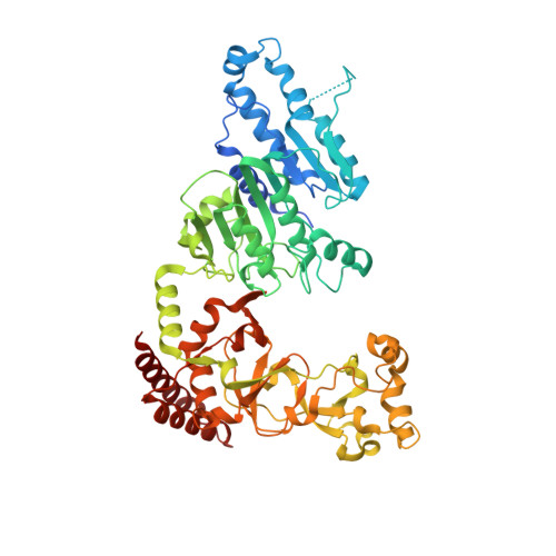
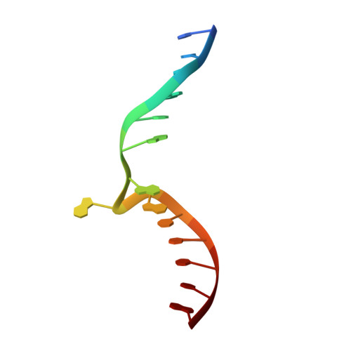
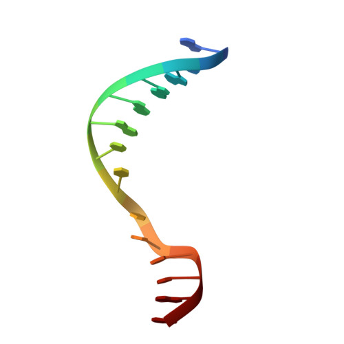
    Antivirulence agents targeting endospore-transmitted Clostridioides difficile infections are urgently needed. C. difficile- specific DNA adenine methyltransferase (CamA) is required for efficient sporulation and affects persistence in the colon. The active site of CamA is conserved and closely resembles those of hundreds of related S -adenosyl-l-methionine (SAM)-dependent methyltransferases, which makes the design of selective inhibitors more challenging. We explored the solvent-exposed edge of the SAM adenosine moiety and systematically designed 42 analogs of adenosine carrying substituents at the C6-amino group (N6) of adenosine. We compare the inhibitory properties and binding affinity of these diverse compounds and present the crystal structures of CamA in complex with 14 of them in the presence of substrate DNA. The most potent of these inhibitors, compound 39 (IC 50 ∼ 0.4 μM and K D ∼ 0.2 μM), is selective for CamA against closely related bacterial and mammalian DNA and RNA adenine methyltransferases, protein lysine and arginine methyltransferases, and human adenosine receptors.


  • Organizational Affiliation

    Department of Epigenetics and Molecular Carcinogenesis, University of Texas MD Anderson Cancer Center, Houston, Texas 77030, United States.


Macromolecules

Find similar proteins by:  (by identity cutoff)  |  3D Structure
Entity ID: 1
MoleculeChains Sequence LengthOrganismDetailsImage
Site-specific DNA-methyltransferase (adenine-specific)
A, B, C
578Clostridioides difficile 630Mutation(s): 0 
Gene Names: CD630_27580
EC: 2.1.1.72
UniProt
Find proteins for Q183J3 (Clostridioides difficile (strain 630))
Explore Q183J3 
Go to UniProtKB:  Q183J3
Entity Groups  
Sequence Clusters30% Identity50% Identity70% Identity90% Identity95% Identity100% Identity
UniProt GroupQ183J3
Sequence Annotations
Expand
  • Reference Sequence

Find similar nucleic acids by:  Sequence   |   3D Structure  

Entity ID: 2
MoleculeChains LengthOrganismImage
DNA Strand 1
D, F, H
14Clostridioides difficile
Sequence Annotations
Expand
  • Reference Sequence

Find similar nucleic acids by:  Sequence   |   3D Structure  

Entity ID: 3
MoleculeChains LengthOrganismImage
DNA Strand 2
E, G, I
14Clostridioides difficile
Sequence Annotations
Expand
  • Reference Sequence
Small Molecules
Ligands 3 Unique
IDChains Name / Formula / InChI Key2D Diagram3D Interactions
Q8L (Subject of Investigation/LOI)
Query on Q8L

Download Ideal Coordinates CCD File 
N [auth A],
U [auth B],
X [auth C]
Piclidenoson
C18 H19 I N6 O4
HUJXGQILHAUCCV-MOROJQBDSA-N
EDO
Query on EDO

Download Ideal Coordinates CCD File 
L [auth A],
M [auth A],
R [auth B],
S [auth B],
T [auth B]
1,2-ETHANEDIOL
C2 H6 O2
LYCAIKOWRPUZTN-UHFFFAOYSA-N
K
Query on K

Download Ideal Coordinates CCD File 
J [auth A]
K [auth A]
O [auth B]
P [auth B]
Q [auth B]
J [auth A],
K [auth A],
O [auth B],
P [auth B],
Q [auth B],
V [auth C],
W [auth C]
POTASSIUM ION
K
NPYPAHLBTDXSSS-UHFFFAOYSA-N
Experimental Data & Validation

Experimental Data

  • Method: X-RAY DIFFRACTION
  • Resolution: 2.78 Å
  • R-Value Free: 0.227 
  • R-Value Work: 0.184 
  • R-Value Observed: 0.186 
  • Space Group: P 21 21 21
Unit Cell:
Length ( Å )Angle ( ˚ )
a = 81.326α = 90
b = 161.634β = 90
c = 229.602γ = 90
Software Package:
Software NamePurpose
SERGUIdata collection
PHENIXrefinement
HKL-2000data reduction
HKL-2000data scaling
PHENIXphasing

Structure Validation

View Full Validation Report


Ligand Structure Quality Assessment 


Entry History & Funding Information

Deposition Data


Funding OrganizationLocationGrant Number
National Institutes of Health/National Institute of General Medical Sciences (NIH/NIGMS)United StatesGM049245-23

Revision History  (Full details and data files)

  • Version 1.0: 2023-01-11
    Type: Initial release
  • Version 1.1: 2023-01-18
    Changes: Structure summary
  • Version 1.2: 2023-01-25
    Changes: Database references
  • Version 1.3: 2023-10-25
    Changes: Data collection, Refinement description