


Crystal structure of Dihydropteridine reductase/oxygen-insensitive NAD(P)H nitroreductase from Klebsiella pneumoniae
Lovell, S., Liu, L., Seibold, S., Battaile, K.P.To be published.
Experimental Data Snapshot
Entity ID: 1 | |||||
|---|---|---|---|---|---|
| Molecule | Chains | Sequence Length | Organism | Details | Image |
| Dihydropteridine reductase/oxygen-insensitive NAD(P)H nitroreductase | 225 | Klebsiella pneumoniae | Mutation(s): 0 Gene Names: nfnB, KPN_00553, KPHS_29700 | 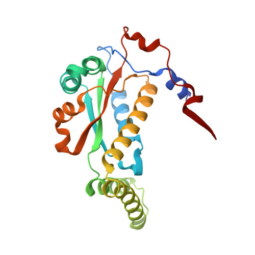 | |
UniProt | |||||
Find proteins for A6T5Y2 (Klebsiella pneumoniae subsp. pneumoniae (strain ATCC 700721 / MGH 78578)) Explore A6T5Y2 Go to UniProtKB: A6T5Y2 | |||||
Entity Groups | |||||
| Sequence Clusters | 30% Identity50% Identity70% Identity90% Identity95% Identity100% Identity | ||||
| UniProt Group | A6T5Y2 | ||||
Sequence AnnotationsExpand | |||||
| |||||
| Ligands 3 Unique | |||||
|---|---|---|---|---|---|
| ID | Chains | Name / Formula / InChI Key | 2D Diagram | 3D Interactions | |
| FMN (Subject of Investigation/LOI) Query on FMN | E [auth A], I [auth B], N [auth C], S [auth D] | FLAVIN MONONUCLEOTIDE C17 H21 N4 O9 P FVTCRASFADXXNN-SCRDCRAPSA-N |  | ||
| PO4 Query on PO4 | F [auth A], J [auth B], O [auth C], T [auth D] | PHOSPHATE ION O4 P NBIIXXVUZAFLBC-UHFFFAOYSA-K |  | ||
| GOL Query on GOL | G [auth A] H [auth A] K [auth B] L [auth B] M [auth B] | GLYCEROL C3 H8 O3 PEDCQBHIVMGVHV-UHFFFAOYSA-N |  | ||
| Length ( Å ) | Angle ( ˚ ) |
|---|---|
| a = 56.826 | α = 90 |
| b = 56.826 | β = 90 |
| c = 257.568 | γ = 120 |
| Software Name | Purpose |
|---|---|
| XDS | data reduction |
| Aimless | data scaling |
| PHASER | phasing |
| PHENIX | refinement |
| PDB_EXTRACT | data extraction |
| Funding Organization | Location | Grant Number |
|---|---|---|
| National Institutes of Health/National Institute Of Allergy and Infectious Diseases (NIH/NIAID) | United States | HHSN272201700059C |
| National Institutes of Health/Office of the Director | United States | S10OD030394 |
RCSB PDB (citation) is hosted by
RCSB PDB is a member of the

