7ZF0

Crystal structure of UGT85B1 from Sorghum bicolor in complex with UDP and p-hydroxymandelonitrile


Experimental Data Snapshot

  • Method: X-RAY DIFFRACTION
  • Resolution: 1.50 Å
  • R-Value Free: 0.210 
  • R-Value Work: 0.189 
  • R-Value Observed: 0.190 

wwPDB Validation   3D Report Full Report


Ligand Structure Quality Assessment 


This is version 1.3 of the entry. See complete history


Literature

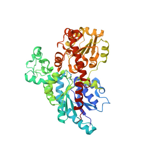
Structure-guided engineering of key amino acids in UGT85B1 controlling substrate and stereo-specificity in aromatic cyanogenic glucoside biosynthesis.

Del Giudice, R.Putkaradze, N.Dos Santos, B.M.Hansen, C.C.Crocoll, C.Motawia, M.S.Fredslund, F.Laursen, T.Welner, D.H.

(2022) Plant J 111: 1539-1549

  • DOI: https://doi.org/10.1111/tpj.15904
  • Primary Citation of Related Structures:  
    7ZER, 7ZF0

  • PubMed Abstract: 

    Cyanogenic glucosides are important defense molecules in plants with useful biological activities in animals. Their last biosynthetic step consists of a glycosylation reaction that confers stability and increases structural diversity and is catalyzed by the UDP-dependent glycosyltransferases (UGTs) of glycosyltransferase family 1. These versatile enzymes have large and varied substrate scopes, and the structure-function relationships controlling scope and specificity remain poorly understood. Here, we report substrate-bound crystal structures and rational engineering of substrate and stereo-specificities of UGT85B1 from Sorghum bicolor involved in biosynthesis of the cyanogenic glucoside dhurrin. Substrate specificity was shifted from the natural substrate (S)-p-hydroxymandelonitrile to (S)-mandelonitrile by combining a mutation to abolish hydrogen bonding to the p-hydroxyl group with a mutation to provide steric hindrance at the p-hydroxyl group binding site (V132A/Q225W). Further, stereo-specificity was shifted from (S) to (R) by substituting four rationally chosen residues within 6 Å of the nitrile group (M312T/A313T/H408F/G409A). These activities were compared to two other UGTs involved in the biosynthesis of aromatic cyanogenic glucosides in Prunus dulcis (almond) and Eucalyptus cladocalyx. Together, these studies enabled us to pinpoint factors that drive substrate and stereo-specificities in the cyanogenic glucoside biosynthetic UGTs. The structure-guided engineering of the functional properties of UGT85B1 enhances our understanding of the evolution of UGTs involved in the biosynthesis of cyanogenic glucosides and will enable future engineering efforts towards new biotechnological applications.


  • Organizational Affiliation

    Plant Biochemistry, Department of Plant and Environmental Sciences, University of Copenhagen, Thorvaldsensvej 40, DK-1871, Copenhagen, Denmark.


Macromolecules
Find similar proteins by:  (by identity cutoff)  |  3D Structure
Entity ID: 1
MoleculeChains Sequence LengthOrganismDetailsImage
Cyanohydrin beta-glucosyltransferase498Sorghum bicolorMutation(s): 0 
Gene Names: UGT85B1HMNGT
EC: 2.4.1.85
UniProt
Find proteins for Q9SBL1 (Sorghum bicolor)
Explore Q9SBL1 
Go to UniProtKB:  Q9SBL1
Entity Groups  
Sequence Clusters30% Identity50% Identity70% Identity90% Identity95% Identity100% Identity
UniProt GroupQ9SBL1
Sequence Annotations
Expand
  • Reference Sequence
Small Molecules
Ligands 3 Unique
IDChains Name / Formula / InChI Key2D Diagram3D Interactions
UDP (Subject of Investigation/LOI)
Query on UDP

Download Ideal Coordinates CCD File 
B [auth A]URIDINE-5'-DIPHOSPHATE
C9 H14 N2 O12 P2
XCCTYIAWTASOJW-XVFCMESISA-N
DHR (Subject of Investigation/LOI)
Query on DHR

Download Ideal Coordinates CCD File 
I [auth A](2S)-HYDROXY(4-HYDROXYPHENYL)ETHANENITRILE
C8 H7 N O2
HOOOPXDSCKBLFG-MRVPVSSYSA-N
EDO
Query on EDO

Download Ideal Coordinates CCD File 
C [auth A]
D [auth A]
E [auth A]
F [auth A]
G [auth A]
C [auth A],
D [auth A],
E [auth A],
F [auth A],
G [auth A],
H [auth A],
J [auth A],
K [auth A],
L [auth A]
1,2-ETHANEDIOL
C2 H6 O2
LYCAIKOWRPUZTN-UHFFFAOYSA-N
Experimental Data & Validation

Experimental Data

  • Method: X-RAY DIFFRACTION
  • Resolution: 1.50 Å
  • R-Value Free: 0.210 
  • R-Value Work: 0.189 
  • R-Value Observed: 0.190 
  • Space Group: P 21 21 21
Unit Cell:
Length ( Å )Angle ( ˚ )
a = 76.63α = 90
b = 91.38β = 90
c = 92.75γ = 90
Software Package:
Software NamePurpose
PHENIXrefinement
XDSdata reduction
XDSdata scaling
PHASERphasing

Structure Validation

View Full Validation Report


Ligand Structure Quality Assessment 


Entry History & Funding Information

Deposition Data


Funding OrganizationLocationGrant Number
Novo Nordisk FoundationDenmarkNNF20CC0035580

Revision History  (Full details and data files)

  • Version 1.0: 2022-07-13
    Type: Initial release
  • Version 1.1: 2022-08-24
    Changes: Database references
  • Version 1.2: 2022-09-28
    Changes: Database references
  • Version 1.3: 2024-01-31
    Changes: Data collection, Refinement description