7YM8

Cryo-EM structure of Nb29-alpha1AAR-miniGsq complex bound to oxymetazoline


Experimental Data Snapshot

  • Method: ELECTRON MICROSCOPY
  • Resolution: 2.92 Å
  • Aggregation State: PARTICLE 
  • Reconstruction Method: SINGLE PARTICLE 

wwPDB Validation   3D Report Full Report


This is version 1.0 of the entry. See complete history


Literature

Structural basis of alpha 1A -adrenergic receptor activation and recognition by an extracellular nanobody.

Toyoda, Y.Zhu, A.Kong, F.Shan, S.Zhao, J.Wang, N.Sun, X.Zhang, L.Yan, C.Kobilka, B.K.Liu, X.

(2023) Nat Commun 14: 3655-3655

  • DOI: https://doi.org/10.1038/s41467-023-39310-x
  • Primary Citation of Related Structures:  
    7YM8, 7YMH, 7YMJ

  • PubMed Abstract: 

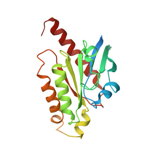
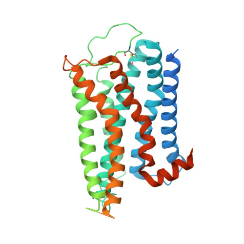
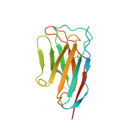
    The α 1A- adrenergic receptor (α 1A AR) belongs to the family of G protein-coupled receptors that respond to adrenaline and noradrenaline. α 1A AR is involved in smooth muscle contraction and cognitive function. Here, we present three cryo-electron microscopy structures of human α 1A AR bound to the endogenous agonist noradrenaline, its selective agonist oxymetazoline, and the antagonist tamsulosin, with resolutions range from 2.9 Å to 3.5 Å. Our active and inactive α 1A AR structures reveal the activation mechanism and distinct ligand binding modes for noradrenaline compared with other adrenergic receptor subtypes. In addition, we identified a nanobody that preferentially binds to the extracellular vestibule of α 1A AR when bound to the selective agonist oxymetazoline. These results should facilitate the design of more selective therapeutic drugs targeting both orthosteric and allosteric sites in this receptor family.


  • Organizational Affiliation

    School of Medicine, Tsinghua University, Beijing, 100084, China. toyoda.yosuke.2r@kyoto-u.ac.jp.


Macromolecules
Find similar proteins by:  (by identity cutoff)  |  3D Structure
Entity ID: 1
MoleculeChains Sequence LengthOrganismDetailsImage
miniGsqA [auth B]246Homo sapiensMutation(s): 0 
Entity Groups  
Sequence Clusters30% Identity50% Identity70% Identity90% Identity95% Identity100% Identity
Sequence Annotations
Expand
  • Reference Sequence
Find similar proteins by:  (by identity cutoff)  |  3D Structure
Entity ID: 2
MoleculeChains Sequence LengthOrganismDetailsImage
alpha1A adrenergic receptorB [auth A]480Homo sapiensMutation(s): 0 
Membrane Entity: Yes 
UniProt & NIH Common Fund Data Resources
Find proteins for D9IEF7 (Enterobacteria phage T4)
Explore D9IEF7 
Go to UniProtKB:  D9IEF7
Find proteins for P35348 (Homo sapiens)
Explore P35348 
Go to UniProtKB:  P35348
PHAROS:  P35348
GTEx:  ENSG00000120907 
Entity Groups  
Sequence Clusters30% Identity50% Identity70% Identity90% Identity95% Identity100% Identity
UniProt GroupsD9IEF7P35348
Sequence Annotations
Expand
  • Reference Sequence
Find similar proteins by:  (by identity cutoff)  |  3D Structure
Entity ID: 3
MoleculeChains Sequence LengthOrganismDetailsImage
nanobody 29C [auth D]144synthetic constructMutation(s): 0 
Entity Groups  
Sequence Clusters30% Identity50% Identity70% Identity90% Identity95% Identity100% Identity
Sequence Annotations
Expand
  • Reference Sequence
Experimental Data & Validation

Experimental Data

  • Method: ELECTRON MICROSCOPY
  • Resolution: 2.92 Å
  • Aggregation State: PARTICLE 
  • Reconstruction Method: SINGLE PARTICLE 

Structure Validation

View Full Validation Report


Entry History & Funding Information

Deposition Data


Funding OrganizationLocationGrant Number
National Natural Science Foundation of China (NSFC)China32122041

Revision History  (Full details and data files)

  • Version 1.0: 2023-07-05
    Type: Initial release