7XJB

Rat-COMT, opicapone,SAM and Mg bond


Experimental Data Snapshot

  • Method: X-RAY DIFFRACTION
  • Resolution: 2.60 Å
  • R-Value Free: 0.194 
  • R-Value Work: 0.162 
  • R-Value Observed: 0.164 

wwPDB Validation   3D Report Full Report


Ligand Structure Quality Assessment 


This is version 1.4 of the entry. See complete history


Literature

Structural and Computational Analyses of the Unique Interactions of Opicapone in the Binding Pocket of Catechol O -Methyltransferase: A Crystallographic Study and Fragment Molecular Orbital Analyses.

Takebe, K.Suzuki, M.Kuwada-Kusunose, T.Shirai, S.Fukuzawa, K.Takamiya, T.Uzawa, N.Iijima, H.

(2023) J Chem Inf Model 63: 4468-4476

  • DOI: https://doi.org/10.1021/acs.jcim.3c00331
  • Primary Citation of Related Structures:  
    7XGI, 7XJB

  • PubMed Abstract: 

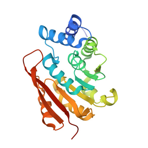
    A third-generation inhibitor of catechol O -methyltransferase (COMT), opicapone ( 1 ), has the 3-nitrocatechol scaffold as do the second-generation inhibitors such as entacapone ( 2 ) and tolcapone ( 3 ), but only 1 can sustainably inhibit COMT activity making it suitable for a once-daily regimen. These improvements should be attributed to the optimized sidechain moiety (oxidopyridyloxadiazolyl group) of 1 substituted at the 5-position of the 3-nitrocatechol ring. We analyzed the role of the sidechain moiety by solving the crystal structures of COMT/ S -adenosylmethionine (SAM)/Mg/ 1 and COMT/ S -adenosylhomocysteine (SAH)/Mg/ 1 complexes. Fragment molecular orbital (FMO) calculations elucidated that the dispersion interaction between the sidechains of Leu 198 and Met 201 on the β6β7-loop and the oxidopyridine ring of 1 were unique and important in both complexes. In contrast, the catechol binding site made a remarkable difference in the sidechain conformation of Lys 144. The ε-amino group of Lys 144 was outside of the catalytic pocket and was replaced by a water molecule in the COMT/SAH/Mg/ 1 complex. No nitrocatechol inhibitor has ever been reported to make a complex with COMT and SAH. Thus, the conformational change of Lys 144 found in the COMT/SAH/Mg/ 1 complex is the first crystallographic evidence that supports the role of Lys 144 as a catalytic base to take out a proton ion from the reaction site to the outside of the enzyme. The fact that 1 generated a complex with SAH and COMT also suggests that 1 could inhibit COMT twofold, as a typical substrate mimic competitive inhibitor and as a product-inhibition enhancer.


  • Organizational Affiliation

    Oral and Maxillofacial Surgery II, Osaka University Graduate School of Dentistry, Suita, Osaka 565-0871, Japan.


Macromolecules
Find similar proteins by:  (by identity cutoff)  |  3D Structure
Entity ID: 1
MoleculeChains Sequence LengthOrganismDetailsImage
Catechol O-methyltransferase
A, B, C, D
223Rattus norvegicusMutation(s): 0 
Gene Names: Comt
EC: 2.1.1.6
UniProt
Find proteins for P22734 (Rattus norvegicus)
Explore P22734 
Go to UniProtKB:  P22734
Entity Groups  
Sequence Clusters30% Identity50% Identity70% Identity90% Identity95% Identity100% Identity
UniProt GroupP22734
Sequence Annotations
Expand
  • Reference Sequence
Small Molecules
Ligands 5 Unique
IDChains Name / Formula / InChI Key2D Diagram3D Interactions
DNI (Subject of Investigation/LOI)
Query on DNI

Download Ideal Coordinates CCD File 
F [auth A],
K [auth B],
O [auth C],
S [auth D]
Opicapone
C15 H10 Cl2 N4 O6
ASOADIZOVZTJSR-UHFFFAOYSA-N
SAM (Subject of Investigation/LOI)
Query on SAM

Download Ideal Coordinates CCD File 
E [auth A],
J [auth B],
N [auth C],
R [auth D]
S-ADENOSYLMETHIONINE
C15 H22 N6 O5 S
MEFKEPWMEQBLKI-FCKMPRQPSA-N
CL
Query on CL

Download Ideal Coordinates CCD File 
H [auth A],
M [auth B],
Q [auth C],
U [auth D],
V [auth D]
CHLORIDE ION
Cl
VEXZGXHMUGYJMC-UHFFFAOYSA-M
MG (Subject of Investigation/LOI)
Query on MG

Download Ideal Coordinates CCD File 
G [auth A],
L [auth B],
P [auth C],
T [auth D]
MAGNESIUM ION
Mg
JLVVSXFLKOJNIY-UHFFFAOYSA-N
NA
Query on NA

Download Ideal Coordinates CCD File 
I [auth A]SODIUM ION
Na
FKNQFGJONOIPTF-UHFFFAOYSA-N
Experimental Data & Validation

Experimental Data

  • Method: X-RAY DIFFRACTION
  • Resolution: 2.60 Å
  • R-Value Free: 0.194 
  • R-Value Work: 0.162 
  • R-Value Observed: 0.164 
  • Space Group: P 43 21 2
Unit Cell:
Length ( Å )Angle ( ˚ )
a = 166.158α = 90
b = 166.158β = 90
c = 125.931γ = 90
Software Package:
Software NamePurpose
PHENIXrefinement
PDB_EXTRACTdata extraction
XDSdata reduction
Aimlessdata scaling
MOLREPphasing

Structure Validation

View Full Validation Report


Ligand Structure Quality Assessment 


Entry History & Funding Information

Deposition Data


Funding OrganizationLocationGrant Number
Japan Society for the Promotion of Science (JSPS)JapanJP15K08034
Japan Agency for Medical Research and Development (AMED)JapanJP18am0101001 (0318)

Revision History  (Full details and data files)

  • Version 1.0: 2023-05-31
    Type: Initial release
  • Version 1.1: 2023-06-21
    Changes: Structure summary
  • Version 1.2: 2023-08-02
    Changes: Database references
  • Version 1.3: 2023-09-27
    Changes: Data collection, Derived calculations, Structure summary
  • Version 1.4: 2023-11-29
    Changes: Refinement description