Structural insights into translation regulation by the THF-II riboswitch.
Xu, L., Xiao, Y., Zhang, J., Fang, X.(2023) Nucleic Acids Res 51: 952-965
- PubMed: 36620887
- DOI: https://doi.org/10.1093/nar/gkac1257
- Primary Citation of Related Structures:
7WI9, 7WIA, 7WIB, 7WIE, 7WIF, 7WII - PubMed Abstract:
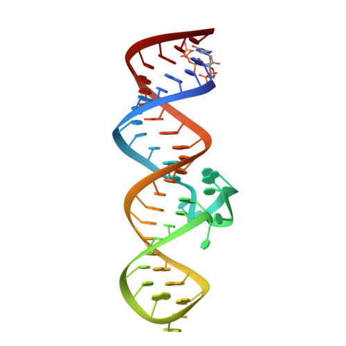
In bacteria, expression of folate-related genes is controlled by the tetrahydrofolate (THF) riboswitch in response to specific binding of THF and its derivatives. Recently, a second class of THF riboswitches, named THF-II, was identified in Gram-negative bacteria, which exhibit distinct architecture from the previously characterized THF-I riboswitches found in Gram-positive bacteria. Here, we present the crystal structures of the ligand-bound THF-II riboswitch from Mesorhizobium loti. These structures exhibit a long rod-like fold stabilized by continuous base pair and base triplet stacking across two helices of P1 and P2 and their interconnecting ligand-bound binding pocket. The pterin moiety of the ligand docks into the binding pocket by forming hydrogen bonds with two highly conserved pyrimidines in J12 and J21, which resembles the hydrogen-bonding pattern at the ligand-binding site FAPK in the THF-I riboswitch. Using small-angle X-ray scattering and isothermal titration calorimetry, we further characterized the riboswitch in solution and reveal that Mg2+ is essential for pre-organization of the binding pocket for efficient ligand binding. RNase H cleavage assay indicates that ligand binding reduces accessibility of the ribosome binding site in the right arm of P1, thus down-regulating the expression of downstream genes. Together, these results provide mechanistic insights into translation regulation by the THF-II riboswitch.
Organizational Affiliation:
Beijing Advanced Innovation Center for Structural Biology, Beijing Frontier Research Center for Biological Structure, School of Life Sciences, Tsinghua University, Beijing 100084, China.