7VEG

Understanding NH-pi interaction between Gln and Phe


Experimental Data Snapshot

  • Method: X-RAY DIFFRACTION
  • Resolution: 1.39 Å
  • R-Value Free: 0.205 
  • R-Value Work: 0.165 
  • R-Value Observed: 0.169 

wwPDB Validation   3D Report Full Report


Ligand Structure Quality Assessment 


This is version 1.2 of the entry. See complete history


Literature

Structural Achievability of an NH-pi Interaction between Gln and Phe in a Crystal Structure of a Collagen-like Peptide.

Zhang, R.Xu, Y.Lan, J.Fan, S.Huang, J.Xu, F.

(2022) Biomolecules 12

  • DOI: https://doi.org/10.3390/biom12101433
  • Primary Citation of Related Structures:  
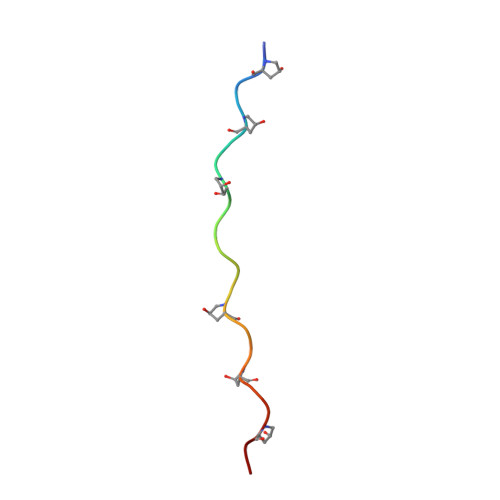
    7VEG

  • PubMed Abstract: 

    NH-π interactions between polar and aromatic residues are well distributed in proteins whose stabilizing effects have been investigated in globular and fibrous proteins. In order to gain structural insights into side chain NH-π interactions, we solved a crystal structure of a collagen-like peptide containing Gln-Phe pairs. The Gln-Phe NH-π interactions were further characterized by quantum calculations, molecular simulations, and structural bioinformatics. The analyses indicated that the NH-π interactions are robust under various solvent conditions, can be distributed either on the protein surface or in its hydrophobic core and can form at a wide range of distances between residues. This study suggested that NH-π interactions can play a versatile role in protein design, including engineering hydrophobic cores, solvent accessible surfaces, and protein-protein interfaces.


  • Organizational Affiliation

    Ministry of Education Key Laboratory of Industrial Biotechnology, School of Biotechnology, Jiangnan University, Wuxi 214122, China.


Macromolecules

Find similar proteins by:  Sequence   |   3D Structure  

Entity ID: 1
MoleculeChains Sequence LengthOrganismDetailsImage
peptide
A, B, C
22Homo sapiensMutation(s): 0 
Entity Groups  
Sequence Clusters30% Identity50% Identity70% Identity90% Identity95% Identity100% Identity
Sequence Annotations
Expand
  • Reference Sequence
Experimental Data & Validation

Experimental Data

  • Method: X-RAY DIFFRACTION
  • Resolution: 1.39 Å
  • R-Value Free: 0.205 
  • R-Value Work: 0.165 
  • R-Value Observed: 0.169 
  • Space Group: P 21 2 21
Unit Cell:
Length ( Å )Angle ( ˚ )
a = 24.093α = 90
b = 28.016β = 90
c = 73.372γ = 90
Software Package:
Software NamePurpose
HKL-2000data reduction
SCALEPACKdata scaling
PHASERphasing
PHENIXrefinement
PDB_EXTRACTdata extraction

Structure Validation

View Full Validation Report


Ligand Structure Quality Assessment 


Entry History & Funding Information

Deposition Data

  • Released Date: 2022-10-19 
  • Deposition Author(s): Fan, S., Xu, F.

Funding OrganizationLocationGrant Number
Not funded--

Revision History  (Full details and data files)

  • Version 1.0: 2022-10-19
    Type: Initial release
  • Version 1.1: 2023-05-03
    Changes: Database references, Refinement description
  • Version 1.2: 2023-11-29
    Changes: Data collection, Refinement description