7U63

Crystal Structure Anti-Oxycodone Antibody HY2-A12 Fab Complexed with Oxycodone


Experimental Data Snapshot

  • Method: X-RAY DIFFRACTION
  • Resolution: 2.30 Å
  • R-Value Free: 0.282 
  • R-Value Work: 0.226 
  • R-Value Observed: 0.229 

wwPDB Validation   3D Report Full Report


Ligand Structure Quality Assessment 


This is version 1.2 of the entry. See complete history


Literature

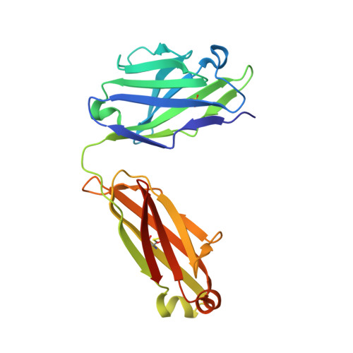
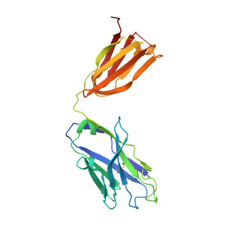
Structures of drug-specific monoclonal antibodies bound to opioids and nicotine reveal a common mode of binding.

Rodarte, J.V.Baehr, C.Hicks, D.Liban, T.L.Weidle, C.Rupert, P.B.Jahan, R.Wall, A.McGuire, A.T.Strong, R.K.Runyon, S.Pravetoni, M.Pancera, M.

(2023) Structure 31: 20-32.e5

  • DOI: https://doi.org/10.1016/j.str.2022.11.008
  • Primary Citation of Related Structures:  
    7U61, 7U62, 7U63, 7U64

  • PubMed Abstract: 

    Opioid-related fatal overdoses have reached epidemic proportions. Because existing treatments for opioid use disorders offer limited long-term protection, accelerating the development of newer approaches is critical. Monoclonal antibodies (mAbs) are an emerging treatment strategy that targets and sequesters selected opioids in the bloodstream, reducing drug distribution across the blood-brain barrier, thus preventing or reversing opioid toxicity. We previously identified a series of murine mAbs with high affinity and selectivity for oxycodone, morphine, fentanyl, and nicotine. To determine their binding mechanism, we used X-ray crystallography to solve the structures of mAbs bound to their respective targets, to 2.2 Å resolution or higher. Structural analysis showed a critical convergent hydrogen bonding mode that is dependent on a glutamic acid residue in the mAbs' heavy chain and a tertiary amine of the ligand. Characterizing drug-mAb complexes represents a significant step toward rational antibody engineering and future manufacturing activities to support clinical evaluation.


  • Organizational Affiliation

    Vaccine and Infectious Disease Division, Fred Hutchinson Cancer Center, Seattle, WA, USA.


Macromolecules
Find similar proteins by:  (by identity cutoff)  |  3D Structure
Entity ID: 1
MoleculeChains Sequence LengthOrganismDetailsImage
HY2-A12 Fab Light ChainA [auth L],
C [auth A],
E [auth C]
215Mus musculusMutation(s): 0 
Entity Groups  
Sequence Clusters30% Identity50% Identity70% Identity90% Identity95% Identity100% Identity
Sequence Annotations
Expand
  • Reference Sequence
Find similar proteins by:  (by identity cutoff)  |  3D Structure
Entity ID: 2
MoleculeChains Sequence LengthOrganismDetailsImage
HY2-A12 Fab Heavy ChainB [auth H],
D [auth B],
F [auth D]
224Mus musculusMutation(s): 0 
Entity Groups  
Sequence Clusters30% Identity50% Identity70% Identity90% Identity95% Identity100% Identity
Sequence Annotations
Expand
  • Reference Sequence
Experimental Data & Validation

Experimental Data

  • Method: X-RAY DIFFRACTION
  • Resolution: 2.30 Å
  • R-Value Free: 0.282 
  • R-Value Work: 0.226 
  • R-Value Observed: 0.229 
  • Space Group: P 1
Unit Cell:
Length ( Å )Angle ( ˚ )
a = 67.624α = 69.13
b = 67.989β = 67.6
c = 85.623γ = 68.64
Software Package:
Software NamePurpose
PHENIXrefinement
SCALEPACKdata scaling
PDB_EXTRACTdata extraction
HKL-2000data reduction
PHASERphasing

Structure Validation

View Full Validation Report


Ligand Structure Quality Assessment 


Entry History & Funding Information

Deposition Data


Funding OrganizationLocationGrant Number
National Institutes of Health/National Institute on Drug Abuse (NIH/NIDA)United StatesUG3 DA047711

Revision History  (Full details and data files)

  • Version 1.0: 2023-01-11
    Type: Initial release
  • Version 1.1: 2023-01-18
    Changes: Database references
  • Version 1.2: 2023-10-25
    Changes: Data collection, Refinement description