7Q6X

OleP mutant S240Y in complex with 6DEB


Experimental Data Snapshot

  • Method: X-RAY DIFFRACTION
  • Resolution: 2.70 Å
  • R-Value Free: 0.268 
  • R-Value Work: 0.204 
  • R-Value Observed: 0.207 

wwPDB Validation   3D Report Full Report


Ligand Structure Quality Assessment 


This is version 1.3 of the entry. See complete history


Literature

Point Mutations at a Key Site Alter the Cytochrome P450 OleP Structural Dynamics.

Montemiglio, L.C.Gugole, E.Freda, I.Exertier, C.D'Auria, L.Chen, C.G.Nardi, A.N.Cerutti, G.Parisi, G.D'Abramo, M.Savino, C.Vallone, B.

(2021) Biomolecules 12

  • DOI: https://doi.org/10.3390/biom12010055
  • Primary Citation of Related Structures:  
    7Q6R, 7Q6X, 7Q89

  • PubMed Abstract: 

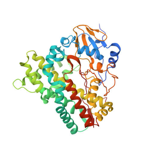
    Substrate binding to the cytochrome P450 OleP is coupled to a large open-to-closed transition that remodels the active site, minimizing its exposure to the external solvent. When the aglycone substrate binds, a small empty cavity is formed between the I and G helices, the BC loop, and the substrate itself, where solvent molecules accumulate mediating substrate-enzyme interactions. Herein, we analyzed the role of this cavity in substrate binding to OleP by producing three mutants (E89Y, G92W, and S240Y) to decrease its volume. The crystal structures of the OleP mutants in the closed state bound to the aglycone 6DEB showed that G92W and S240Y occupied the cavity, providing additional contact points with the substrate. Conversely, mutation E89Y induces a flipped-out conformation of this amino acid side chain, that points towards the bulk, increasing the empty volume. Equilibrium titrations and molecular dynamic simulations indicate that the presence of a bulky residue within the cavity impacts the binding properties of the enzyme, perturbing the conformational space explored by the complexes. Our data highlight the relevance of this region in OleP substrate binding and suggest that it represents a key substrate-protein contact site to consider in the perspective of redirecting its activity towards alternative compounds.


  • Organizational Affiliation

    Institute of Molecular Biology and Pathology, CNR c/o Department of Biochemical Sciences "A. Rossi Fanelli", University of Rome, Sapienza, P.le A. Moro 5, 00185 Rome, Italy.


Macromolecules
Find similar proteins by:  (by identity cutoff)  |  3D Structure
Entity ID: 1
MoleculeChains Sequence LengthOrganismDetailsImage
Cytochrome P-450
A, B, C, D, E
A, B, C, D, E, F
406Streptomyces antibioticusMutation(s): 1 
Gene Names: oleP
UniProt
Find proteins for Q59819 (Streptomyces antibioticus)
Explore Q59819 
Go to UniProtKB:  Q59819
Entity Groups  
Sequence Clusters30% Identity50% Identity70% Identity90% Identity95% Identity100% Identity
UniProt GroupQ59819
Sequence Annotations
Expand
  • Reference Sequence
Small Molecules
Ligands 3 Unique
IDChains Name / Formula / InChI Key2D Diagram3D Interactions
HEM
Query on HEM

Download Ideal Coordinates CCD File 
G [auth A]
L [auth B]
N [auth C]
Q [auth D]
T [auth E]
G [auth A],
L [auth B],
N [auth C],
Q [auth D],
T [auth E],
W [auth F]
PROTOPORPHYRIN IX CONTAINING FE
C34 H32 Fe N4 O4
KABFMIBPWCXCRK-RGGAHWMASA-L
DEB
Query on DEB

Download Ideal Coordinates CCD File 
H [auth A]
M [auth B]
O [auth C]
R [auth D]
U [auth E]
H [auth A],
M [auth B],
O [auth C],
R [auth D],
U [auth E],
X [auth F]
6-DEOXYERYTHRONOLIDE B
C21 H38 O6
HQZOLNNEQAKEHT-IBBGRPSASA-N
FMT
Query on FMT

Download Ideal Coordinates CCD File 
I [auth A]
J [auth A]
K [auth A]
P [auth C]
S [auth D]
I [auth A],
J [auth A],
K [auth A],
P [auth C],
S [auth D],
V [auth E]
FORMIC ACID
C H2 O2
BDAGIHXWWSANSR-UHFFFAOYSA-N
Experimental Data & Validation

Experimental Data

  • Method: X-RAY DIFFRACTION
  • Resolution: 2.70 Å
  • R-Value Free: 0.268 
  • R-Value Work: 0.204 
  • R-Value Observed: 0.207 
  • Space Group: C 1 2 1
Unit Cell:
Length ( Å )Angle ( ˚ )
a = 248.624α = 90
b = 110.355β = 129.63
c = 160.046γ = 90
Software Package:
Software NamePurpose
REFMACrefinement
XDSdata reduction
SCALAdata scaling
MOLREPphasing

Structure Validation

View Full Validation Report


Ligand Structure Quality Assessment 


Entry History & Funding Information

Deposition Data


Funding OrganizationLocationGrant Number
Not funded--

Revision History  (Full details and data files)

  • Version 1.0: 2022-01-26
    Type: Initial release
  • Version 1.1: 2022-02-09
    Changes: Database references
  • Version 1.2: 2022-04-06
    Changes: Advisory, Derived calculations, Structure summary
  • Version 1.3: 2024-01-31
    Changes: Data collection, Refinement description