7PGB

NaV_Ae1/Sp1CTD_pore-SAT09 complex


Experimental Data Snapshot

  • Method: X-RAY DIFFRACTION
  • Resolution: 3.60 Å
  • R-Value Free: 0.232 
  • R-Value Work: 0.207 
  • R-Value Observed: 0.208 

wwPDB Validation   3D Report Full Report


Ligand Structure Quality Assessment 


This is version 1.3 of the entry. See complete history


Literature

Quaternary structure independent folding of voltage-gated ion channel pore domain subunits.

Arrigoni, C.Lolicato, M.Shaya, D.Rohaim, A.Findeisen, F.Fong, L.K.Colleran, C.M.Dominik, P.Kim, S.S.Schuermann, J.P.DeGrado, W.F.Grabe, M.Kossiakoff, A.A.Minor Jr., D.L.

(2022) Nat Struct Mol Biol 29: 537-548

  • DOI: https://doi.org/10.1038/s41594-022-00775-x
  • Primary Citation of Related Structures:  
    7PG8, 7PGB, 7PGF, 7PGG, 7PGH, 7PGI

  • PubMed Abstract: 

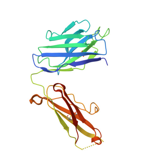
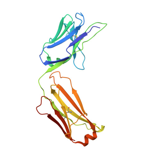
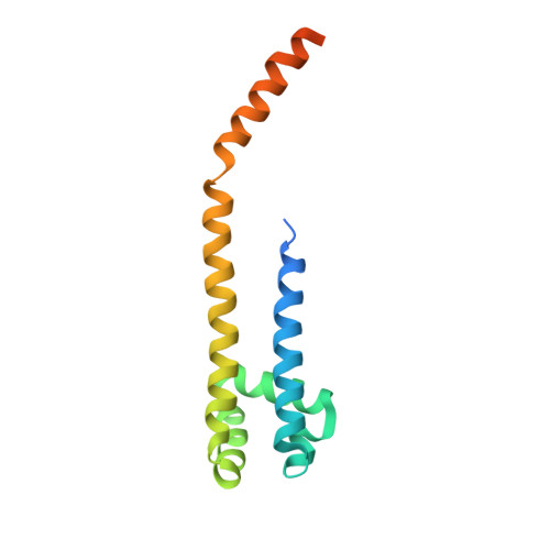
    Every voltage-gated ion channel (VGIC) has a pore domain (PD) made from four subunits, each comprising an antiparallel transmembrane helix pair bridged by a loop. The extent to which PD subunit structure requires quaternary interactions is unclear. Here, we present crystal structures of a set of bacterial voltage-gated sodium channel (BacNa V ) 'pore only' proteins that reveal a surprising collection of non-canonical quaternary arrangements in which the PD tertiary structure is maintained. This context-independent structural robustness, supported by molecular dynamics simulations, indicates that VGIC-PD tertiary structure is independent of quaternary interactions. This fold occurs throughout the VGIC superfamily and in diverse transmembrane and soluble proteins. Strikingly, characterization of PD subunit-binding Fabs indicates that non-canonical quaternary PD conformations can occur in full-length VGICs. Together, our data demonstrate that the VGIC-PD is an autonomously folded unit. This property has implications for VGIC biogenesis, understanding functional states, de novo channel design, and VGIC structural origins.


  • Organizational Affiliation

    Cardiovascular Research Institute, University of California, San Francisco, CA, USA.


Macromolecules
Find similar proteins by:  (by identity cutoff)  |  3D Structure
Entity ID: 1
MoleculeChains Sequence LengthOrganismDetailsImage
SAT09 fab fragment, heavy chain234Homo sapiensMutation(s): 0 
Entity Groups  
Sequence Clusters30% Identity50% Identity70% Identity90% Identity95% Identity100% Identity
Sequence Annotations
Expand
  • Reference Sequence
Find similar proteins by:  (by identity cutoff)  |  3D Structure
Entity ID: 2
MoleculeChains Sequence LengthOrganismDetailsImage
SAT09 fab fragment, light chain215Homo sapiensMutation(s): 0 
Entity Groups  
Sequence Clusters30% Identity50% Identity70% Identity90% Identity95% Identity100% Identity
Sequence Annotations
Expand
  • Reference Sequence
Find similar proteins by:  (by identity cutoff)  |  3D Structure
Entity ID: 3
MoleculeChains Sequence LengthOrganismDetailsImage
Ion transport protein,Voltage-gated sodium channel143Alkalilimnicola ehrlichii MLHE-1Ruegeria pomeroyi DSS-3
This entity is chimeric
Mutation(s): 0 
Gene Names: Mlg_0322SPO0030
Membrane Entity: Yes 
UniProt
Find proteins for Q0ABW0 (Alkalilimnicola ehrlichii (strain ATCC BAA-1101 / DSM 17681 / MLHE-1))
Explore Q0ABW0 
Go to UniProtKB:  Q0ABW0
Find proteins for F7IVA8 (Ruegeria pomeroyi (strain ATCC 700808 / DSM 15171 / DSS-3))
Explore F7IVA8 
Go to UniProtKB:  F7IVA8
Entity Groups  
Sequence Clusters30% Identity50% Identity70% Identity90% Identity95% Identity100% Identity
UniProt GroupsQ0ABW0F7IVA8
Sequence Annotations
Expand
  • Reference Sequence
Small Molecules
Ligands 10 Unique
IDChains Name / Formula / InChI Key2D Diagram3D Interactions
LHG
Query on LHG

Download Ideal Coordinates CCD File 
FC [auth p]
GA [auth H]
HB [auth X]
LA [auth A]
MC [auth f]
FC [auth p],
GA [auth H],
HB [auth X],
LA [auth A],
MC [auth f],
OC [auth i],
PB [auth a],
RA [auth R],
WB [auth m],
ZA [auth U]
1,2-DIPALMITOYL-PHOSPHATIDYL-GLYCEROLE
C38 H75 O10 P
BIABMEZBCHDPBV-MPQUPPDSSA-N
PE4
Query on PE4

Download Ideal Coordinates CCD File 
IA [auth H]2-{2-[2-(2-{2-[2-(2-ETHOXY-ETHOXY)-ETHOXY]-ETHOXY}-ETHOXY)-ETHOXY]-ETHOXY}-ETHANOL
C16 H34 O8
PJWQOENWHPEPKI-UHFFFAOYSA-N
BHC
Query on BHC

Download Ideal Coordinates CCD File 
SC [auth h]BENZENE HEXACARBOXYLIC ACID
C12 H6 O12
YDSWCNNOKPMOTP-UHFFFAOYSA-N
1PE
Query on 1PE

Download Ideal Coordinates CCD File 
CC [auth l],
MA [auth A],
SA [auth R],
UC [auth h]
PENTAETHYLENE GLYCOL
C10 H22 O6
JLFNLZLINWHATN-UHFFFAOYSA-N
R16
Query on R16

Download Ideal Coordinates CCD File 
GC [auth p]HEXADECANE
C16 H34
DCAYPVUWAIABOU-UHFFFAOYSA-N
D12
Query on D12

Download Ideal Coordinates CCD File 
JC [auth o],
OB [auth a],
ZC [auth e]
DODECANE
C12 H26
SNRUBQQJIBEYMU-UHFFFAOYSA-N
4NB
Query on 4NB

Download Ideal Coordinates CCD File 
AC [auth l]
BB [auth W]
EA [auth H]
EC [auth p]
FA [auth H]
AC [auth l],
BB [auth W],
EA [auth H],
EC [auth p],
FA [auth H],
FB [auth X],
GB [auth X],
HC [auth o],
IC [auth o],
JB [auth Z],
KB [auth Z],
LB [auth Z],
LC [auth f],
NA [auth C],
NC [auth i],
OA [auth C],
PC [auth h],
QA [auth R],
QC [auth h],
RB [auth d],
SB [auth d],
TA [auth T],
UA [auth T],
VA [auth T],
VB [auth m],
WC [auth e],
XB [auth l],
XC [auth e],
YA [auth U],
YB [auth l],
YC [auth e],
ZB [auth l]
4-NITROBENZOIC ACID
C7 H5 N O4
OTLNPYWUJOZPPA-UHFFFAOYSA-N
OCT
Query on OCT

Download Ideal Coordinates CCD File 
AB [auth U]
BC [auth l]
CB [auth W]
HA [auth H]
IB [auth X]
AB [auth U],
BC [auth l],
CB [auth W],
HA [auth H],
IB [auth X],
JA [auth c],
KC [auth o],
PA [auth C],
QB [auth a],
RC [auth h],
WA [auth T]
N-OCTANE
C8 H18
TVMXDCGIABBOFY-UHFFFAOYSA-N
GOL
Query on GOL

Download Ideal Coordinates CCD File 
TC [auth h]GLYCEROL
C3 H8 O3
PEDCQBHIVMGVHV-UHFFFAOYSA-N
LNK
Query on LNK

Download Ideal Coordinates CCD File 
AD [auth e]
BD [auth e]
CD [auth e]
DB [auth W]
DC [auth l]
AD [auth e],
BD [auth e],
CD [auth e],
DB [auth W],
DC [auth l],
EB [auth W],
KA [auth c],
MB [auth Z],
NB [auth Z],
TB [auth d],
UB [auth d],
VC [auth h],
XA [auth T]
PENTANE
C5 H12
OFBQJSOFQDEBGM-UHFFFAOYSA-N
Experimental Data & Validation

Experimental Data

  • Method: X-RAY DIFFRACTION
  • Resolution: 3.60 Å
  • R-Value Free: 0.232 
  • R-Value Work: 0.207 
  • R-Value Observed: 0.208 
  • Space Group: P 43
Unit Cell:
Length ( Å )Angle ( ˚ )
a = 200.865α = 90
b = 200.865β = 90
c = 327.727γ = 90
Software Package:
Software NamePurpose
Cootmodel building
PHENIXrefinement
XDSdata reduction
Aimlessdata scaling
PHASERphasing

Structure Validation

View Full Validation Report


Ligand Structure Quality Assessment 


Entry History & Funding Information

Deposition Data


Funding OrganizationLocationGrant Number
National Institutes of Health/National Heart, Lung, and Blood Institute (NIH/NHLBI)United States--
National Institutes of Health/National Institute on Deafness and Other Communication Disorders (NIH/NIDCD)United States--
National Institutes of Health/National Institute of General Medical Sciences (NIH/NIGMS)United States--

Revision History  (Full details and data files)

  • Version 1.0: 2022-06-08
    Type: Initial release
  • Version 1.1: 2022-06-15
    Changes: Database references
  • Version 1.2: 2022-06-29
    Changes: Database references
  • Version 1.3: 2024-01-31
    Changes: Data collection, Refinement description