7OUB

High resolution structure of Alpha-1-acid glycoprotein bound to potent anti-tumour compound UCN-01


Experimental Data Snapshot

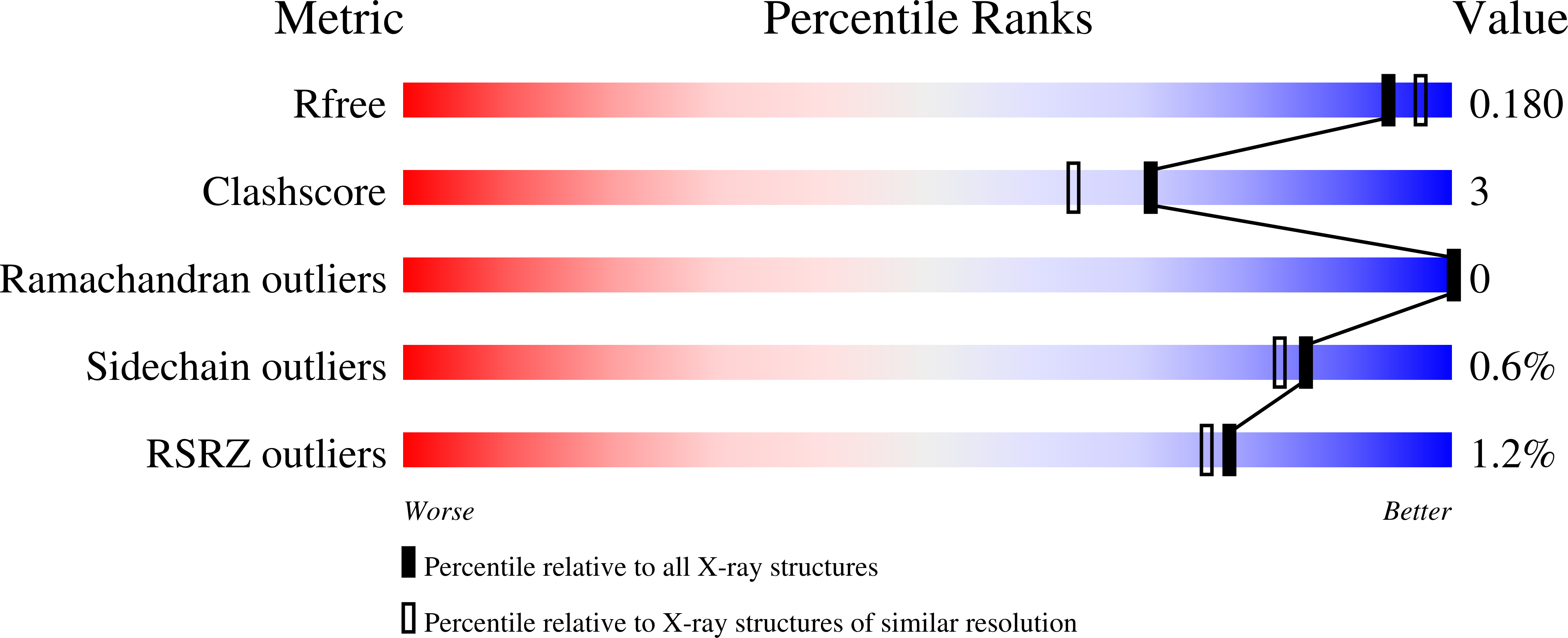
  • Method: X-RAY DIFFRACTION
  • Resolution: 1.82 Å
  • R-Value Free: 0.177 
  • R-Value Work: 0.156 
  • R-Value Observed: 0.157 

wwPDB Validation   3D Report Full Report


This is version 1.2 of the entry. See complete history


Literature

The structural basis for high affinity binding of alpha 1-acid glycoprotein to the potent antitumor compound UCN-01.

Landin, E.J.B.Williams, C.Ryan, S.A.Bochel, A.Akter, N.Redfield, C.Sessions, R.B.Dedi, N.Taylor, R.J.Crump, M.P.

(2021) J Biol Chem 297: 101392-101392

  • DOI: https://doi.org/10.1016/j.jbc.2021.101392
  • Primary Citation of Related Structures:  
    7OUB

  • PubMed Abstract: 

    The α1-acid glycoprotein (AGP) is an abundant blood plasma protein with important immunomodulatory functions coupled to endogenous and exogenous ligand-binding properties. Its affinity for many drug-like structures, however, means AGP can have a significant effect on the pharmokinetics and pharmacodynamics of numerous small molecule therapeutics. Staurosporine, and its hydroxylated forms UCN-01 and UCN-02, are kinase inhibitors that have been investigated at length as antitumour compounds. Despite their potency, these compounds display poor pharmokinetics due to binding to both AGP variants, AGP1 and AGP2. The recent renewed interest in UCN-01 as a cytostatic protective agent prompted us to solve the structure of the AGP2-UCN-01 complex by X-ray crystallography, revealing for the first time the precise binding mode of UCN-01. The solution NMR suggests AGP2 undergoes a significant conformational change upon ligand binding, but also that it uses a common set of sidechains with which it captures key groups of UCN-01 and other small molecule ligands. We anticipate that this structure and the supporting NMR data will facilitate rational redesign of small molecules that could evade AGP and therefore improve tissue distribution.


  • Organizational Affiliation

    School of Chemistry, University of Bristol, Bristol, UK.


Macromolecules
Find similar proteins by:  (by identity cutoff)  |  3D Structure
Entity ID: 1
MoleculeChains Sequence LengthOrganismDetailsImage
Alpha-1-acid glycoprotein 2A [auth B]191Homo sapiensMutation(s): 1 
Gene Names: ORM2AGP2
UniProt & NIH Common Fund Data Resources
Find proteins for P19652 (Homo sapiens)
Explore P19652 
Go to UniProtKB:  P19652
PHAROS:  P19652
GTEx:  ENSG00000228278 
Entity Groups  
Sequence Clusters30% Identity50% Identity70% Identity90% Identity95% Identity100% Identity
UniProt GroupP19652
Sequence Annotations
Expand
  • Reference Sequence
Small Molecules
Ligands 1 Unique
IDChains Name / Formula / InChI Key2D Diagram3D Interactions
UCN (Subject of Investigation/LOI)
Query on UCN

Download Ideal Coordinates CCD File 
B
7-HYDROXYSTAUROSPORINE
C28 H26 N4 O4
PBCZSGKMGDDXIJ-HQCWYSJUSA-N
Experimental Data & Validation

Experimental Data

  • Method: X-RAY DIFFRACTION
  • Resolution: 1.82 Å
  • R-Value Free: 0.177 
  • R-Value Work: 0.156 
  • R-Value Observed: 0.157 
  • Space Group: P 61
Unit Cell:
Length ( Å )Angle ( ˚ )
a = 88.27α = 90
b = 88.27β = 90
c = 53.84γ = 120
Software Package:
Software NamePurpose
PHENIXrefinement
PDB_EXTRACTdata extraction
XDSdata reduction
DIALSdata scaling
PHASERphasing

Structure Validation

View Full Validation Report


Entry History & Funding Information

Deposition Data


Funding OrganizationLocationGrant Number
Biotechnology and Biological Sciences Research Council (BBSRC)United KingdomBB/L01386X/1
Commonwealth Scholarship Commission (United Kingdom)United KingdomBDCS-2017-50

Revision History  (Full details and data files)

  • Version 1.0: 2021-12-01
    Type: Initial release
  • Version 1.1: 2021-12-22
    Changes: Database references
  • Version 1.2: 2024-01-31
    Changes: Data collection, Refinement description